IPLab:Lab 3:Healed Myocardial Infarction
Clinical Summary
This 57-year-old white female with a 22 year history of insulin-dependent diabetes was admitted to the hospital 10 hours prior to death complaining of shortness of breath. Three months before, she had begun to experience progressive weakness and for the previous 3 weeks she noticed increasing dyspnea on exertion and worsening of a chronic cough. Despite appropriate care she developed an intractable arrhythmia and could not be resuscitated.
Autopsy showed a 340-gram heart with extensive transmural reddish discoloration of the anterolateral portion of the myocardium of the left ventricle. There was severe atherosclerotic narrowing of all coronary arteries and an acute thrombus in the left anterior descending. The lungs showed pulmonary edema and early bronchopneumonia.
Images
-
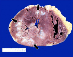 This is a gross photograph of a heart with areas of old healed myocardial infarction (scars) outlined by arrows.
 -
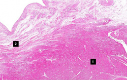 This is a low-power photomicrograph of a healed myocardial infarction with a fibrous scar. Remaining normal tissue is on the right (1) and the fibrous connective tissue scar is on the left (2).
 -
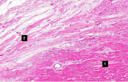 This is a higher-power photomicrograph of the healed myocardial infarction with a fibrous scar. The normal tissue is on the right (1) and the fibrous connective tissue scar is on the upper left (2).
 -
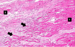 This is a higher-power photomicrograph of a healed myocardial infarction with a fibrous scar. Remaining normal tissue is at the right (1) and the fibrous connective tissue scar is at the left (2). Note the presence of occasional hypereosinophilic myocytes (arrows) indicating recent acute ischemic injury to this region of old scar tissue.
 -
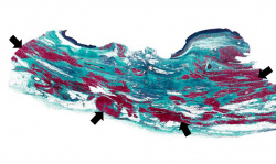 This is a low-power photomicrograph of myocardial tissue stained with a trichrome stain to demonstrate fibrous connective tissue (blue-green). The section shows the mature fibrous connective tissue from this heart with an old healed infarct. Some viable myocardial tissue remains (arrows).
 -
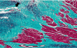 This is a photomicrograph of a trichrome-stained section of heart containing an old healed myocardial infarction. The scar is composed of mature fibrous connective tissue (blue-green) and some remaining viable myocytes (red). A small focus of adipose tissue can be seen in the scar (arrow).


Virtual Microscopy
Early (Inflammatory Response)
Late (Replacement Fibrosis)
Study Questions
Additional Resources
Reference
Journal Articles
- Yuan S, Liu Y, Zhu L. Vascular complications of diabetes mellitus. Clin Exp Pharmacol Physiol 1999 Dec;26(12):977-8.
Images
Related IPLab Cases
- Lab 1: Heart: Myocardial Infarction (Coagulative Necrosis)
- Lab 3: Heart: Acute Myocardial Infarction
- Lab 4: Heart: Mural Thrombus
- Lab 4: Coronary Artery: Thrombosis
- Lab 4: Lung: Pulmonary Congestion and Edema