IPLab:Lab 12:Burns
Jump to navigation
Jump to search
Clinical Summary
This 45-year-old female was involved in a house fire which killed her husband. The patient had approximately 80% body surface area burns including 1st, 2nd, and 3rd degree burns. The patient was intubated and given aggressive fluid resuscitation and ventilatory support, but she remained hypotensive and became progressively hypoxemic and hypercapnic until she died approximately eight hours after the fire.
Carbonaceous material was noted in the throat and posterior pharynx as well as in the trachea and main stem bronchi. The liver was soft, yellow and greasy. Results of premortem blood work showed an elevated blood alcohol level.
Images
-
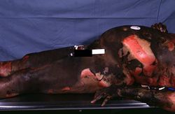 This photograph taken at autopsy demonstrates the severity of the surface burns on this patient.
 -
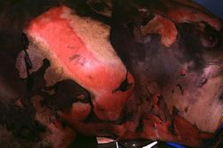 This closer view shows the burned skin peeling off to expose the underlying tissue. The various depths of the burn can be appreciated by the color and character of the underlying tissue.
 -
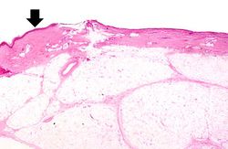 This photomicrograph of the burned skin depicts an area of third degree burn. There is some residual epidermis (arrow) but in the majority of this section the epidermis is gone. Also note the severe subcutaneous edema.
 -
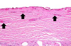 This high-power photomicrograph shows the denuded surface of the skin with thrombosed blood vessels (arrows) and necrosis of the dermis.
 -
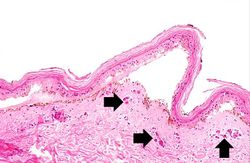 This medium-power photomicrograph of the burned skin demonstrates blister formation. The vessels in the dermis are congested (arrows).
 -
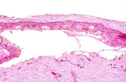 This high-power photomicrograph of the burned skin shows the blister and the thrombosed vessels in the dermis.
 -
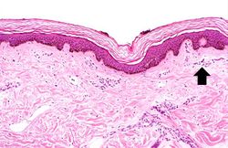 This photomicrograph of the burned skin depicts an area of first degree burn. Note that there are no blisters and no damage to the dermis. There is mild damage to the superficial epidermis and some hyperemia (arrow).
 -
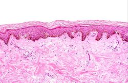 This medium-power photomicrograph of the burned skin shows the mild damage to the superficial layers of the epidermis.
 -
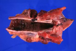 This photograph demonstrates the black carbonaceous material in the trachea.
 -
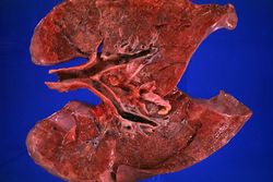 This photograph of the lung again shows the black carbonaceous material in the trachea as well as in the main stem bronchi. The lungs are mildly congested and hyperemic. The patient lived for less than 8 hours after the burn injury so there was not enough time to develop a full-blown pneumonia.
 -
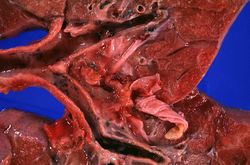 This closer view of the lung shows the black carbonaceous material in the trachea as well as in the main stem bronchi.
 -
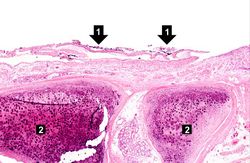 This photomicrograph of the trachea shows sloughing of the tracheal epithelium and the black carbonaceous material contained therein (1). This degree of tracheal epithelial damage is indicative of severe inhalation injury. The tracheal cartilage rings can be seen in cross-section (2).
 -
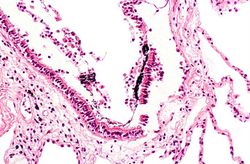 The damage to the tracheal epithelium and the black carbonaceous material is present down to the level of the terminal bronchioles.


Study Questions
Additional Resources
Reference
- eMedicine Medical Library: Thermal Burns
- eMedicine Medical Library: Emergent Management of Thermal Burns
- Merck Manual: Burns
Journal Articles
- Fukuzuka K, Rosenberg JJ, Gaines GC, Edwards CK 3rd, Clare-Salzler M, MacKay SL, Moldawer LL, Copeland EM 3rd, Mozingo DW. Caspase-3-dependent organ apoptosis early after burn injury. Ann Surg 1999 Jun;229(6):851-8; discussion 858-9.
- Robert L. Sheridan, M.D.Fire-Related Inhalation Injury. NEJM 2016 Aug;375:464-469.
Images
| |||||