File:IPLab8HBV6.jpg
Revision as of 02:49, 21 August 2013 by Seung Park (talk | contribs) (This is a photomicrograph of a liver section from another case of hepatitis B. In this H&E-stained section, the typical "ground glass" appearance of the hepatocytes can be appreciated (arrows).)
No higher resolution available.
IPLab8HBV6.jpg (680 × 450 pixels, file size: 70 KB, MIME type: image/jpeg)
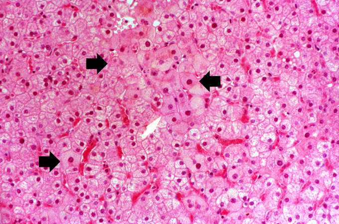
This is a photomicrograph of a liver section from another case of hepatitis B. In this H&E-stained section, the typical "ground glass" appearance of the hepatocytes can be appreciated (arrows).
File history
Click on a date/time to view the file as it appeared at that time.
| Date/Time | Thumbnail | Dimensions | User | Comment | |
|---|---|---|---|---|---|
| current | 02:49, 21 August 2013 | | 680 × 450 (70 KB) | Seung Park (talk | contribs) | This is a photomicrograph of a liver section from another case of hepatitis B. In this H&E-stained section, the typical "ground glass" appearance of the hepatocytes can be appreciated (arrows). |
- You cannot overwrite this file.
File usage
The following page links to this file: