File:IPLab8HSVGlossitis2.jpg
No higher resolution available.
IPLab8HSVGlossitis2.jpg (674 × 450 pixels, file size: 90 KB, MIME type: image/jpeg)
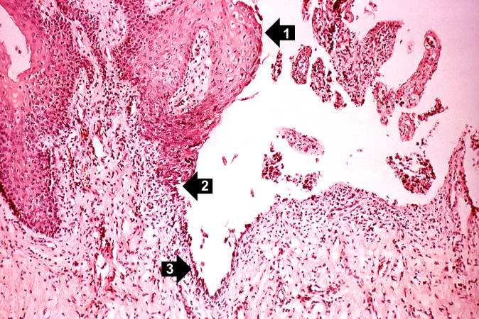
This higher-power photomicrograph shows the epithelium (1), the edge of the ulcer (2), and the ulcerated epithelium (3). There is an inflammatory exudate at the base of the ulcer and some necrotic cells where the epithelium once was present.
File history
Click on a date/time to view the file as it appeared at that time.
| Date/Time | Thumbnail | Dimensions | User | Comment | |
|---|---|---|---|---|---|
| current | 21:28, 20 August 2013 | | 674 × 450 (90 KB) | Seung Park (talk | contribs) | This higher-power photomicrograph shows the epithelium (1), the edge of the ulcer (2), and the ulcerated epithelium (3). There is an inflammatory exudate at the base of the ulcer and some necrotic cells where the epithelium once was present. |
You cannot overwrite this file.
File usage
The following page uses this file:
