IPLab:Lab 7:Osteosarcoma
Images
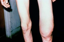
This is a photograph of the patient prior to surgery. Note the marked swelling of the knee.
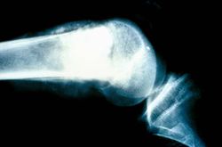
This is a radiograph showing the tumor in the distal femur.
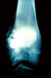
This is another view of the tumor in the distal femur.
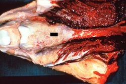
This is a gross photograph of the surgical specimen with tissue dissected away to demonstrate the tumor mass.
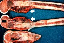
These are cut sections of the distal femur containing the tumor. The periosteal involvement is evident from this picture (arrows).
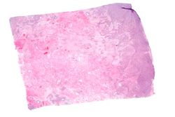
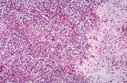
This is a low-power photomicrograph of decalcified histologic section from this tumor. Note the blue color (cell nuclei stain blue) of much of this section indicating the increased cellularity of the tumor.
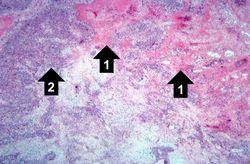
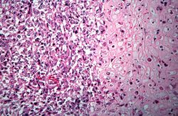
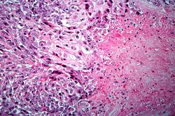
This is a higher-power photomicrograph of decalcified histologic section from this tumor. There are areas of osteoid (1) and cellular areas (2).
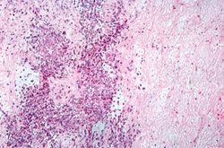
This is a high-power photomicrograph of decalcified histologic section showing the cellularity of the tumor.
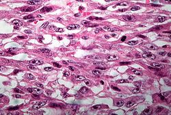
This high-power photomicrograph demonstrates the cellular growth pattern. Note that the cells are fusiform and they grow in sheets.
This high-power photomicrograph demonstrates the growth pattern and the cell morphology.
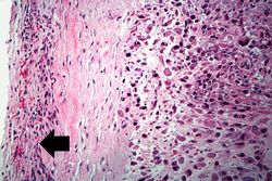
This is a high-power photomicrograph of the tumor cell morphology and the periosteum (arrow).
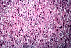
This high-power photomicrograph of the tumor demonstrates the fusiform morphology of the cells. Note the marked variability in size and staining intensity of the nuclei.
This is a high-power photomicrograph of the tumor demonstrating the anaplastic cell morphology.
This is a high-power photomicrograph of the tumor demonstrating the anaplastic cell morphology.
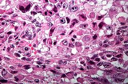
This is a high-power photomicrograph of the tumor demonstrating the anaplastic cell morphology and multiple mitotic figures (arrows).


