IPLab:Lab 7:Carcinoid
Images
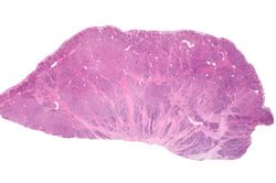
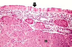
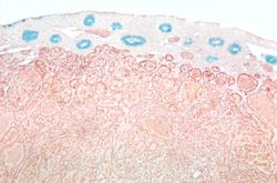
This is a low-power photomicrograph of the surgical specimen showing basophilic and eosinophilic areas delimiting areas of tumor infiltration.
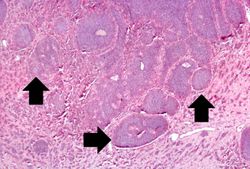
This is a higher-power photomicrograph of the surgical specimen showing nests of tumor cells (arrows).
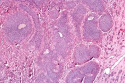
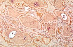
This is a high-power photomicrograph of the surgical specimen showing the tumor's growth pattern--cells form discrete islands, trabeculae, and glands.
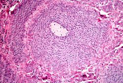
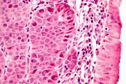
This is a high-power photomicrograph of the surgical specimen showing the cellular morphology. The tumor cells are monotonously similar with scant, pink, granular cytoplasm and a round-to-oval stippled nucleus. As in most carcinoid tumors, there is minimal variation in cell and nuclear size, and mitoses are infrequent or absent.
This is a low-power photomicrograph of one of the subcutaneous masses in the cecum. Note that the mucosa (1) is virtually normal and the tumor cells are in the submucosa (2).
This is a higher-power photomicrograph of the previous section showing the intact mucosa (right) and the submucosal carcinoid tumor.
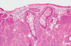
This is a low-power photomicrograph of another one of the subcutaneous masses in the cecum. The mucosa is normal and the tumor cells are in the submucosa.
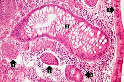
This is a higher-power photomicrograph of the previous section showing intact mucosa (1), a gland (2), and the submucosal carcinoid tumor cells (3).
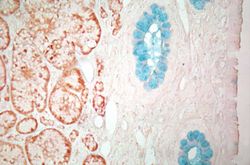
This is a low-power photomicrograph of a section of cecum containing tumor stained to demonstrate the secretory granules in these tumor cells (brown-colored stain). The blue color is the mucin in the glands just under the mucosal surface.
This is a higher-power view of the previous section stained with a silver stain to delineate carcinoid tumor cells (brown) and a mucin stain (blue) to stain the glands.
This is a high-power view of the same section stained with a silver stain to delineate carcinoid tumor cells (brown).


