File:IPLab7Carcinoid8.jpg
No higher resolution available.
IPLab7Carcinoid8.jpg (677 × 450 pixels, file size: 81 KB, MIME type: image/jpeg)
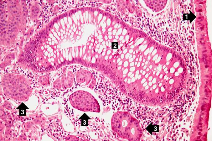
This is a higher-power photomicrograph of the previous section showing intact mucosa (1), a gland (2), and the submucosal carcinoid tumor cells (3).
File history
Click on a date/time to view the file as it appeared at that time.
| Date/Time | Thumbnail | Dimensions | User | Comment | |
|---|---|---|---|---|---|
| current | 21:08, 20 August 2013 | | 677 × 450 (81 KB) | Seung Park (talk | contribs) | This is a higher-power photomicrograph of the previous section showing intact mucosa (1), a gland (2), and the submucosal carcinoid tumor cells (3). |
You cannot overwrite this file.
File usage
There are no pages that use this file.
