File:IPLab7Carcinoid3.jpg
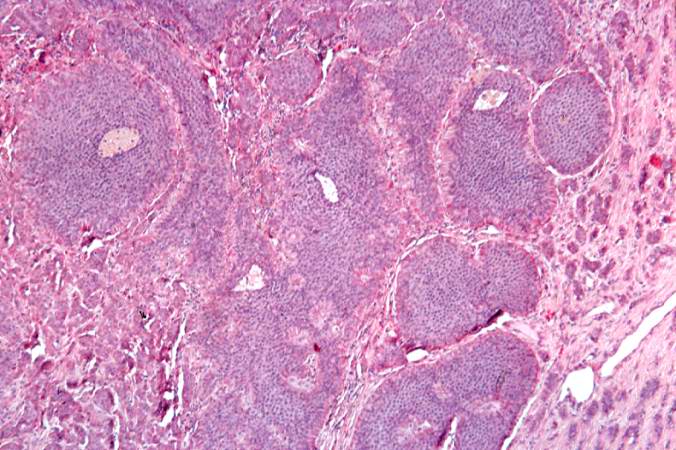
Revision as of 02:06, 21 August 2013 by Seung Park (talk | contribs) (This is a high-power photomicrograph of the surgical specimen showing the tumor's growth pattern--cells form discrete islands, trabeculae, and glands.)
No higher resolution available.
IPLab7Carcinoid3.jpg (676 × 450 pixels, file size: 72 KB, MIME type: image/jpeg)
This is a high-power photomicrograph of the surgical specimen showing the tumor's growth pattern--cells form discrete islands, trabeculae, and glands.
File history
Click on a date/time to view the file as it appeared at that time.
| Date/Time | Thumbnail | Dimensions | User | Comment | |
|---|---|---|---|---|---|
| current | 02:06, 21 August 2013 | | 676 × 450 (72 KB) | Seung Park (talk | contribs) | This is a high-power photomicrograph of the surgical specimen showing the tumor's growth pattern--cells form discrete islands, trabeculae, and glands. |
- You cannot overwrite this file.
File usage
There are no pages that link to this file.