File:IPLab7Fibroadenoma6.jpg
No higher resolution available.
IPLab7Fibroadenoma6.jpg (684 × 450 pixels, file size: 61 KB, MIME type: image/jpeg)
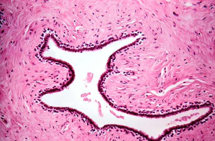
This is a high magnification of the fibroadenoma showing the dense stroma of the tumor surrounding the irregularly shaped duct. The ducts are lined by two cell layers, one of cuboidal, two columnar cells (inner layer) and an outer layer of flattened cells with hyperchromatic nuclei (myoepithelial cells).
File history
Click on a date/time to view the file as it appeared at that time.
| Date/Time | Thumbnail | Dimensions | User | Comment | |
|---|---|---|---|---|---|
| current | 20:28, 20 August 2013 | | 684 × 450 (61 KB) | Seung Park (talk | contribs) | This is a high magnification of the fibroadenoma showing the dense stroma of the tumor surrounding the irregularly shaped duct. The ducts are lined by two cell layers, one of cuboidal, two columnar cells (inner layer) and an outer layer of flattened ce... |
You cannot overwrite this file.
File usage
There are no pages that use this file.
