File:IPLab7Adenoma4.jpg
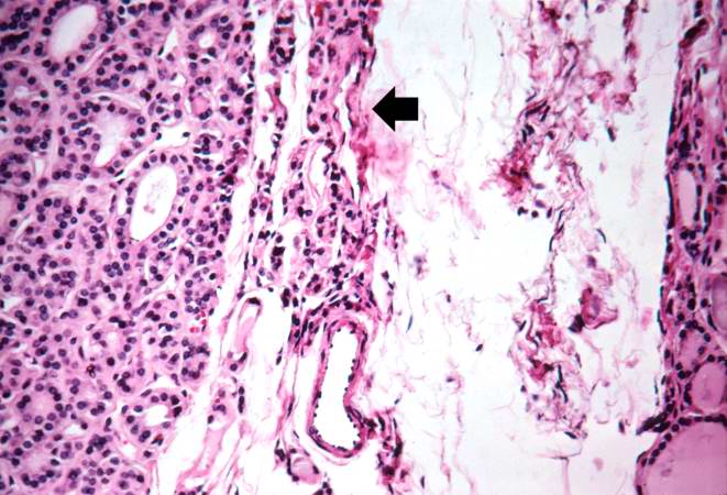
Revision as of 01:23, 21 August 2013 by Seung Park (talk | contribs) (This photomicrograph demonstrates the densely packed follicular pattern in the adenoma (left) and the larger colloid-filled follicles of the normal thyroid (right). An area of compressed thyroid is present adjacent to the adenoma (arrows).)
No higher resolution available.
IPLab7Adenoma4.jpg (661 × 450 pixels, file size: 61 KB, MIME type: image/jpeg)
This photomicrograph demonstrates the densely packed follicular pattern in the adenoma (left) and the larger colloid-filled follicles of the normal thyroid (right). An area of compressed thyroid is present adjacent to the adenoma (arrows).
File history
Click on a date/time to view the file as it appeared at that time.
| Date/Time | Thumbnail | Dimensions | User | Comment | |
|---|---|---|---|---|---|
| current | 01:23, 21 August 2013 | | 661 × 450 (61 KB) | Seung Park (talk | contribs) | This photomicrograph demonstrates the densely packed follicular pattern in the adenoma (left) and the larger colloid-filled follicles of the normal thyroid (right). An area of compressed thyroid is present adjacent to the adenoma (arrows). |
- You cannot overwrite this file.
File usage
There are no pages that link to this file.