File:IPLab7Adenoma2.jpg
Revision as of 01:22, 21 August 2013 by Seung Park (talk | contribs) (This is a higher-power view of the border between the tumor mass and the adjacent thyroid tissue. Note that the mass has compressed the adjacent normal thyroid tissue (arrow). Also note the different morphology between the adenoma (very cellular, dense...)
No higher resolution available.
IPLab7Adenoma2.jpg (674 × 450 pixels, file size: 72 KB, MIME type: image/jpeg)
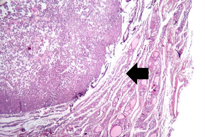
This is a higher-power view of the border between the tumor mass and the adjacent thyroid tissue. Note that the mass has compressed the adjacent normal thyroid tissue (arrow). Also note the different morphology between the adenoma (very cellular, dense follicles, little or no colloid) and the adjacent normal thyroid (larger follicles, colloid).
File history
Click on a date/time to view the file as it appeared at that time.
| Date/Time | Thumbnail | Dimensions | User | Comment | |
|---|---|---|---|---|---|
| current | 01:22, 21 August 2013 | | 674 × 450 (72 KB) | Seung Park (talk | contribs) | This is a higher-power view of the border between the tumor mass and the adjacent thyroid tissue. Note that the mass has compressed the adjacent normal thyroid tissue (arrow). Also note the different morphology between the adenoma (very cellular, dense... |
- You cannot overwrite this file.
File usage
There are no pages that link to this file.