IPLab:Lab 6:Multiple Myeloma
Images
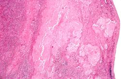
This is a low-power photomicrograph of the mediastinal mass. The mass is encapsulated and contains cellular areas (blue) and areas of pale red material.
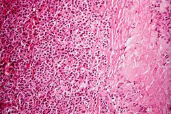
This higher-power photomicrograph shows the junction between an amorphous hyaline-appearing area (amyloid) on the right and cellular areas (plasmacytoid cells) on the left.
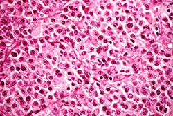
This high-power photomicrograph demonstrates the cells that make up this tissue. These cells resemble plasma cells and are the malignant cell of multiple myeloma.
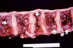
This is a photograph of the vertebral column from this patient at autopsy. Notice the collapsed vertebra (1). There are multiple variably-sized white nodules (2) within the bone marrow. These are accumulations of malignant plasma cells in this case of multiple myeloma.


