File:IPLab6Amyloid4.jpg
Revision as of 21:34, 20 August 2013 by Seung Park (talk | contribs) (This is a low-power photomicrograph of liver tissue from this case. Note the eosinophilic hyaline material (1) present within and between the hepatic tissue (2). There is marked distortion of lobular architecture by the amyloid.)
No higher resolution available.
IPLab6Amyloid4.jpg (681 × 450 pixels, file size: 97 KB, MIME type: image/jpeg)
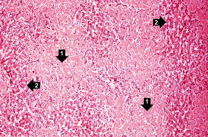
This is a low-power photomicrograph of liver tissue from this case. Note the eosinophilic hyaline material (1) present within and between the hepatic tissue (2). There is marked distortion of lobular architecture by the amyloid.
File history
Click on a date/time to view the file as it appeared at that time.
| Date/Time | Thumbnail | Dimensions | User | Comment | |
|---|---|---|---|---|---|
| current | 21:34, 20 August 2013 | | 681 × 450 (97 KB) | Seung Park (talk | contribs) | This is a low-power photomicrograph of liver tissue from this case. Note the eosinophilic hyaline material (1) present within and between the hepatic tissue (2). There is marked distortion of lobular architecture by the amyloid. |
- You cannot overwrite this file.
File usage
The following page links to this file: