File:IPLab6RA9.jpg
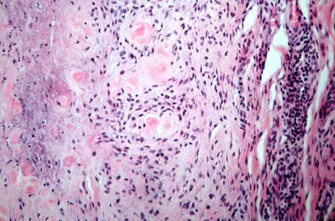
Revision as of 18:08, 19 August 2013 by Seung Park (talk | contribs) (This is a high-power photomicrograph of the mononuclear cells which surround the central area of necrosis. The focal accumulations of fibrinoid material are clearly visible. Lymphocytes are present in the extreme right of this image.)
No higher resolution available.
IPLab6RA9.jpg (682 × 450 pixels, file size: 63 KB, MIME type: image/jpeg)
This is a high-power photomicrograph of the mononuclear cells which surround the central area of necrosis. The focal accumulations of fibrinoid material are clearly visible. Lymphocytes are present in the extreme right of this image.
File history
Click on a date/time to view the file as it appeared at that time.
| Date/Time | Thumbnail | Dimensions | User | Comment | |
|---|---|---|---|---|---|
| current | 18:08, 19 August 2013 | | 682 × 450 (63 KB) | Seung Park (talk | contribs) | This is a high-power photomicrograph of the mononuclear cells which surround the central area of necrosis. The focal accumulations of fibrinoid material are clearly visible. Lymphocytes are present in the extreme right of this image. |
- You cannot overwrite this file.
File usage
The following page links to this file: