File:IPLab4Thromboembolus7.jpg
Revision as of 16:48, 19 August 2013 by Seung Park (talk | contribs) (This is a gross photograph of an infarcted testis. Because of the anatomy of the blood supply to the testis, torsion or the blood vessels often leads to venous occlusion (due to compression of the thin walled veins) but not arterial occlusion. Thus, bl...)
No higher resolution available.
IPLab4Thromboembolus7.jpg (679 × 450 pixels, file size: 64 KB, MIME type: image/jpeg)
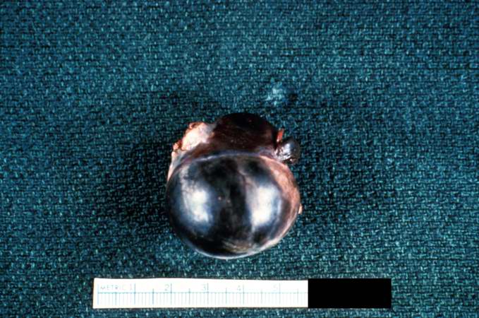
This is a gross photograph of an infarcted testis. Because of the anatomy of the blood supply to the testis, torsion or the blood vessels often leads to venous occlusion (due to compression of the thin walled veins) but not arterial occlusion. Thus, blood still flows into the testis but it can’t get out! This leads to hypoxia and eventually to hemorrhagic necrosis.
An occlusion is a blockage.
File history
Click on a date/time to view the file as it appeared at that time.
| Date/Time | Thumbnail | Dimensions | User | Comment | |
|---|---|---|---|---|---|
| current | 16:48, 19 August 2013 | | 679 × 450 (64 KB) | Seung Park (talk | contribs) | This is a gross photograph of an infarcted testis. Because of the anatomy of the blood supply to the testis, torsion or the blood vessels often leads to venous occlusion (due to compression of the thin walled veins) but not arterial occlusion. Thus, bl... |
- You cannot overwrite this file.
File usage
The following page links to this file: