File:IPLab4Thromboembolus5.jpg
Revision as of 16:48, 19 August 2013 by Seung Park (talk | contribs) (This is a photomicrograph of the wall of the pulmonary artery (1) containing the thromboembolus. In this case the artery wall looks normal. If this was a thrombus instead of a thromboembolus, you would expect to see some damage in the artery wall that ...)
No higher resolution available.
IPLab4Thromboembolus5.jpg (683 × 450 pixels, file size: 66 KB, MIME type: image/jpeg)
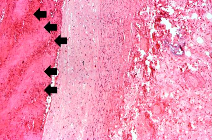
This is a photomicrograph of the wall of the pulmonary artery (1) containing the thromboembolus. In this case the artery wall looks normal. If this was a thrombus instead of a thromboembolus, you would expect to see some damage in the artery wall that would have initiated the thrombus. Note the lines of Zahn in the thromboembolus (arrows).
A thrombus is a solid mass resulting from the aggregation of blood constituents within the vascular system.
File history
Click on a date/time to view the file as it appeared at that time.
| Date/Time | Thumbnail | Dimensions | User | Comment | |
|---|---|---|---|---|---|
| current | 16:48, 19 August 2013 | | 683 × 450 (66 KB) | Seung Park (talk | contribs) | This is a photomicrograph of the wall of the pulmonary artery (1) containing the thromboembolus. In this case the artery wall looks normal. If this was a thrombus instead of a thromboembolus, you would expect to see some damage in the artery wall that ... |
- You cannot overwrite this file.
File usage
The following page links to this file: