File:IPLab4Thrombosis2.jpg
Revision as of 16:35, 19 August 2013 by Seung Park (talk | contribs) (This is a low-power photomicrograph of thrombosed coronary artery. The thrombus (1) completely occludes the vessel. Note the layering of the thrombus. The fibrous cap is ruptured (arrow) and there is hemorrhage into the atherosclerotic plaque. Note the...)
No higher resolution available.
IPLab4Thrombosis2.jpg (651 × 450 pixels, file size: 42 KB, MIME type: image/jpeg)
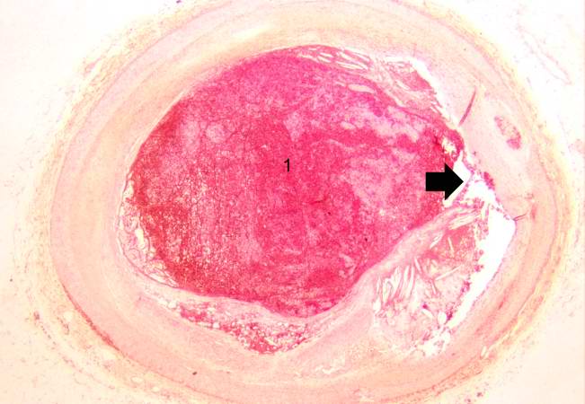
This is a low-power photomicrograph of thrombosed coronary artery. The thrombus (1) completely occludes the vessel. Note the layering of the thrombus. The fibrous cap is ruptured (arrow) and there is hemorrhage into the atherosclerotic plaque. Note the cholesterol crystals in the plaque.
A thrombus is a solid mass resulting from the aggregation of blood constituents within the vascular system.
File history
Click on a date/time to view the file as it appeared at that time.
| Date/Time | Thumbnail | Dimensions | User | Comment | |
|---|---|---|---|---|---|
| current | 16:35, 19 August 2013 | | 651 × 450 (42 KB) | Seung Park (talk | contribs) | This is a low-power photomicrograph of thrombosed coronary artery. The thrombus (1) completely occludes the vessel. Note the layering of the thrombus. The fibrous cap is ruptured (arrow) and there is hemorrhage into the atherosclerotic plaque. Note the... |
- You cannot overwrite this file.
File usage
There are no pages that link to this file.