File:IPLab4MuralThrombus4.jpg
Revision as of 16:28, 19 August 2013 by Seung Park (talk | contribs) (This is a high-power photomicrograph of the border zone between the thrombus (1) and the endocardium (2). In this region there is less inflammation at the border zone.)
No higher resolution available.
IPLab4MuralThrombus4.jpg (677 × 450 pixels, file size: 65 KB, MIME type: image/jpeg)
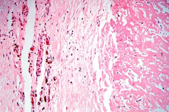
This is a high-power photomicrograph of the border zone between the thrombus (1) and the endocardium (2). In this region there is less inflammation at the border zone.
A thrombus is a solid mass resulting from the aggregation of blood constituents within the vascular system.
File history
Click on a date/time to view the file as it appeared at that time.
| Date/Time | Thumbnail | Dimensions | User | Comment | |
|---|---|---|---|---|---|
| current | 16:28, 19 August 2013 | | 677 × 450 (65 KB) | Seung Park (talk | contribs) | This is a high-power photomicrograph of the border zone between the thrombus (1) and the endocardium (2). In this region there is less inflammation at the border zone. |
- You cannot overwrite this file.
File usage
The following page links to this file: