File:IPLab4ChronicPassiveCongestion2.jpg
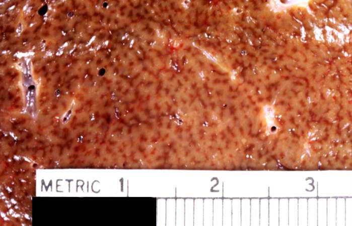
Revision as of 16:19, 19 August 2013 by Seung Park (talk | contribs) (This is a closer view of a cut section of liver demonstrating the pattern of chronic passive congestion. The central vein regions are red and the surrounding hepatic tissue is pale tan-brown.)
No higher resolution available.
IPLab4ChronicPassiveCongestion2.jpg (699 × 450 pixels, file size: 44 KB, MIME type: image/jpeg)
This is a closer view of a cut section of liver demonstrating the pattern of chronic passive congestion. The central vein regions are red and the surrounding hepatic tissue is pale tan-brown.
File history
Click on a date/time to view the file as it appeared at that time.
| Date/Time | Thumbnail | Dimensions | User | Comment | |
|---|---|---|---|---|---|
| current | 16:19, 19 August 2013 | | 699 × 450 (44 KB) | Seung Park (talk | contribs) | This is a closer view of a cut section of liver demonstrating the pattern of chronic passive congestion. The central vein regions are red and the surrounding hepatic tissue is pale tan-brown. |
- You cannot overwrite this file.
File usage
The following page links to this file: