File:IPLab3Bronchopneumonia4.jpg
No higher resolution available.
IPLab3Bronchopneumonia4.jpg (675 × 450 pixels, file size: 94 KB, MIME type: image/jpeg)
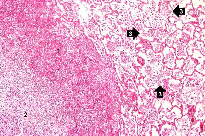
This photomicrograph of the wall of an abscess (1) illustrates liquefaction necrosis in the center of the abscess (2). The remaining lung parenchyma (on the right side of the image) has extensive neutrophilic infiltration into the alveoli (3). This abscess has destroyed a portion of the lung, but other areas of the lung where the normal structure has been retained could recover normal function.
File history
Click on a date/time to view the file as it appeared at that time.
| Date/Time | Thumbnail | Dimensions | User | Comment | |
|---|---|---|---|---|---|
| current | 22:23, 18 August 2013 | | 675 × 450 (94 KB) | Seung Park (talk | contribs) | This photomicrograph of the wall of an abscess (1) illustrates liquefaction necrosis in the center of the abscess (2). The remaining lung parenchyma (on the right side of the image) has extensive neutrophilic infiltration into the alveoli (3). This abs... |
You cannot overwrite this file.
File usage
The following page uses this file:
