File:IPLab3Bronchopneumonia3.jpg
IPLab3Bronchopneumonia3.jpg (469 × 450 pixels, file size: 28 KB, MIME type: image/jpeg)
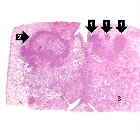
This is a low-power photomicrograph of lung with multiple focal lesions (1) throughout the tissue, some of which have a pale center indicating a loss of parenchymal tissue (2). This is typical of abscess formation in the lung and represents a form of liquefaction necrosis. This pneumonia is bronchopneumonia since the distribution is along the bronchi and the terminal airway distribution throughout the lung. Note that there are some areas of lung which appear relatively normal, having a pale-staining appearance. Other areas, such as in the lower central area (3), are much darker, indicating a heavy cellular infiltrate.
File history
Click on a date/time to view the file as it appeared at that time.
| Date/Time | Thumbnail | Dimensions | User | Comment | |
|---|---|---|---|---|---|
| current | 22:22, 18 August 2013 | | 469 × 450 (28 KB) | Seung Park (talk | contribs) | This is a low-power photomicrograph of lung with multiple focal lesions (1) throughout the tissue, some of which have a pale center indicating a loss of parenchymal tissue (2). This is typical of abscess formation in the lung and represents a form of l... |
You cannot overwrite this file.
File usage
The following page uses this file:
