File:IPLab3LobarPneumonia8.jpg
Revision as of 03:18, 19 August 2013 by Seung Park (talk | contribs) (This is a photomicrograph of alveoli filled with exudate. The alveolar wall outlines (arrows) are barely visible in this section. The alveoli are filled with PMNs, fibrin, and edema fluid. This is a severe acute inflammatory response but the structure ...)
No higher resolution available.
IPLab3LobarPneumonia8.jpg (676 × 450 pixels, file size: 92 KB, MIME type: image/jpeg)
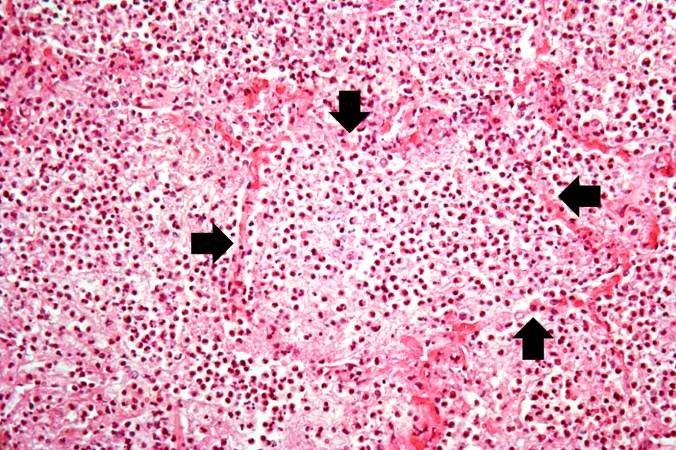
This is a photomicrograph of alveoli filled with exudate. The alveolar wall outlines (arrows) are barely visible in this section. The alveoli are filled with PMNs, fibrin, and edema fluid. This is a severe acute inflammatory response but the structure of the alveoli remains intact. This tissue is able, with proper treatment, to completely resolve this inflammatory response. Since there has not been necrosis of the lung tissue itself (loss of tissue), this lung could completely recover normal function (resolution).
File history
Click on a date/time to view the file as it appeared at that time.
| Date/Time | Thumbnail | Dimensions | User | Comment | |
|---|---|---|---|---|---|
| current | 03:18, 19 August 2013 | | 676 × 450 (92 KB) | Seung Park (talk | contribs) | This is a photomicrograph of alveoli filled with exudate. The alveolar wall outlines (arrows) are barely visible in this section. The alveoli are filled with PMNs, fibrin, and edema fluid. This is a severe acute inflammatory response but the structure ... |
- You cannot overwrite this file.
File usage
The following page links to this file: