File:IPLab3LobarPneumonia3.jpg
No higher resolution available.
IPLab3LobarPneumonia3.jpg (675 × 450 pixels, file size: 55 KB, MIME type: image/jpeg)
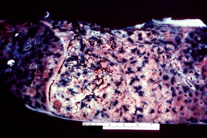
This is a gross photograph of the right lung from the patient in this case. This lung shows complete consolidation with a marked infiltration of neutrophils throughout the tissue giving the lung a whitish discoloration. Note the extensive black pigment in this lung due to anthracosis. This is an advanced case of lobar pneumonia with extensive necrosis which obviously did not resolve, thus resulting in the death of the patient.
File history
Click on a date/time to view the file as it appeared at that time.
| Date/Time | Thumbnail | Dimensions | User | Comment | |
|---|---|---|---|---|---|
| current | 22:17, 18 August 2013 | | 675 × 450 (55 KB) | Seung Park (talk | contribs) | This is a gross photograph of the right lung from the patient in this case. This lung shows complete consolidation with a marked infiltration of neutrophils throughout the tissue giving the lung a whitish discoloration. Note the extensive black pigment... |
You cannot overwrite this file.
File usage
The following page uses this file:
