File:IPLab3LobarPneumonia1.jpg
Revision as of 03:15, 19 August 2013 by Seung Park (talk | contribs) (This is a gross photograph of the lungs from a patient (not the patient from this case) with acute lobar pneumonia. The lung lobe in the upper-right portion of the photograph is affected with pneumonia (arrows). It has a whitish discoloration and appea...)
No higher resolution available.
IPLab3LobarPneumonia1.jpg (680 × 450 pixels, file size: 58 KB, MIME type: image/jpeg)
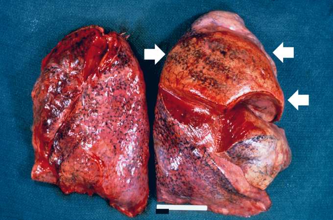
This is a gross photograph of the lungs from a patient (not the patient from this case) with acute lobar pneumonia. The lung lobe in the upper-right portion of the photograph is affected with pneumonia (arrows). It has a whitish discoloration and appears swollen compared to the more pink-staining normal lung lobe in the lower right and left-hand portions of this photograph.
In alcoholics, aspiration pneumonia is common--bacteria enter the lung via aspiration of gastric contents.
File history
Click on a date/time to view the file as it appeared at that time.
| Date/Time | Thumbnail | Dimensions | User | Comment | |
|---|---|---|---|---|---|
| current | 03:15, 19 August 2013 | | 680 × 450 (58 KB) | Seung Park (talk | contribs) | This is a gross photograph of the lungs from a patient (not the patient from this case) with acute lobar pneumonia. The lung lobe in the upper-right portion of the photograph is affected with pneumonia (arrows). It has a whitish discoloration and appea... |
- You cannot overwrite this file.
File usage
The following page links to this file: