File:IPLab13Meningococcemia4.jpg
Revision as of 06:05, 21 August 2013 by Seung Park (talk | contribs) (This photomicrograph of the skin shows thrombi and fibrin clots in small vessels in the dermis. This is indicative of the endothelial damage caused by the Neisseria meningitidis endotoxin. This endotoxin-induced damage to the endothelium of small blood...)
No higher resolution available.
IPLab13Meningococcemia4.jpg (688 × 450 pixels, file size: 75 KB, MIME type: image/jpeg)
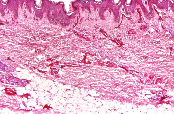
This photomicrograph of the skin shows thrombi and fibrin clots in small vessels in the dermis. This is indicative of the endothelial damage caused by the Neisseria meningitidis endotoxin. This endotoxin-induced damage to the endothelium of small blood vessels throughout the body results in widespread petechiae and purpura.
File history
Click on a date/time to view the file as it appeared at that time.
| Date/Time | Thumbnail | Dimensions | User | Comment | |
|---|---|---|---|---|---|
| current | 06:05, 21 August 2013 | | 688 × 450 (75 KB) | Seung Park (talk | contribs) | This photomicrograph of the skin shows thrombi and fibrin clots in small vessels in the dermis. This is indicative of the endothelial damage caused by the Neisseria meningitidis endotoxin. This endotoxin-induced damage to the endothelium of small blood... |
- You cannot overwrite this file.
File usage
The following page links to this file: