IPLab:Lab 13:Wilms Tumor
Images
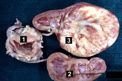
This is a gross photograph of a bladder (1) to which are attached a normal kidney (2) and a kidney with Wilms' tumor (3). A large mass extends from the superior pole of the affected kidney. The renal capsule can be seen extending around this tumor.
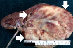
This is a closer view of the kidney with Wilms' tumor (arrows).
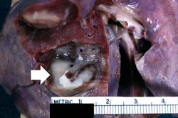
This is a gross photograph of lung from this case demonstrating the metastatic tumor nodule (arrow).
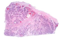
This lowest-power view shows the tumor itself; no tissue is present that can be readily identified as normal kidney. There does appear to be a capsule surrounding the tumor. Eosinophilic bands are seen surrounding basophilic islands of cells. These correspond to the two types of tissue in this tumor--the basophilic cellular component termed "blastema" can be distinguished from less cellular eosinophilic areas.
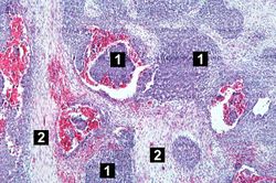
This low-power photomicrograph of tumor shows the two cell types making up this neoplasm. The basophilic cellular component termed "blastema" (1) can be distinguished from less cellular eosinophilic areas with fibroblast-like cells (2).
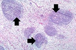
This medium-power photomicrograph of tumor shows again the two cell types making up this neoplasm. There are regions within the blastema where the cells form glands or "tubules" (arrows).
This is another medium-power photomicrograph of the tumor. It demonstrates again the two cell types making up this neoplasm. The glands or "tubules" within the blastema are better developed in this section (arrows).
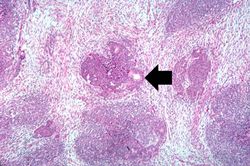
This is a high-power photomicrograph of "tubule" formation within the blastema (arrows).
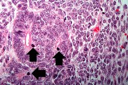
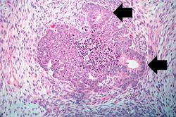
This high-power photomicrograph demonstrates tubule formation within the blastema. Note the numerous mitotic figures (arrows).

This high-power photomicrograph shows the differences in cell morphology between the blastema (1) and the fibroblast type cells (2).


| |||||