File:IPLab13Hyaline7.jpg
Revision as of 05:51, 21 August 2013 by Seung Park (talk | contribs) (This high-power photomicrograph shows an airway with adjacent lung tissue. Some alveoli have hyaline membranes (arrows). There is severe congestion of the interstitium throughout this section.)
No higher resolution available.
IPLab13Hyaline7.jpg (680 × 450 pixels, file size: 93 KB, MIME type: image/jpeg)
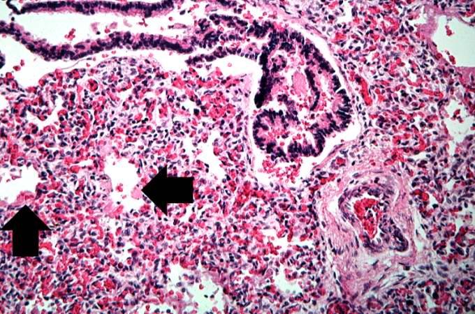
This high-power photomicrograph shows an airway with adjacent lung tissue. Some alveoli have hyaline membranes (arrows). There is severe congestion of the interstitium throughout this section.
File history
Click on a date/time to view the file as it appeared at that time.
| Date/Time | Thumbnail | Dimensions | User | Comment | |
|---|---|---|---|---|---|
| current | 05:51, 21 August 2013 | | 680 × 450 (93 KB) | Seung Park (talk | contribs) | This high-power photomicrograph shows an airway with adjacent lung tissue. Some alveoli have hyaline membranes (arrows). There is severe congestion of the interstitium throughout this section. |
- You cannot overwrite this file.
File usage
The following page links to this file: