File:IPLab13Myelomeningocele5.jpg
Revision as of 05:45, 21 August 2013 by Seung Park (talk | contribs) (This is a higher-power photomicrograph of one of the vertebral bodies from this case. The defect (arrows) in the vertebral body is seen more clearly. The spinal cord is disrupted and there are areas of hemorrhage in this region.)
No higher resolution available.
IPLab13Myelomeningocele5.jpg (679 × 450 pixels, file size: 49 KB, MIME type: image/jpeg)
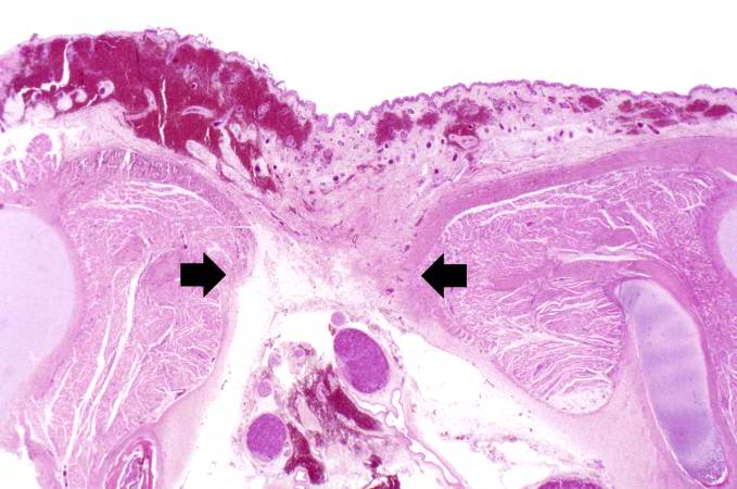
This is a higher-power photomicrograph of one of the vertebral bodies from this case. The defect (arrows) in the vertebral body is seen more clearly. The spinal cord is disrupted and there are areas of hemorrhage in this region.
File history
Click on a date/time to view the file as it appeared at that time.
| Date/Time | Thumbnail | Dimensions | User | Comment | |
|---|---|---|---|---|---|
| current | 05:45, 21 August 2013 | | 679 × 450 (49 KB) | Seung Park (talk | contribs) | This is a higher-power photomicrograph of one of the vertebral bodies from this case. The defect (arrows) in the vertebral body is seen more clearly. The spinal cord is disrupted and there are areas of hemorrhage in this region. |
- You cannot overwrite this file.
File usage
The following page links to this file: