File:IPLab13Myelomeningocele4.jpg
Revision as of 05:45, 21 August 2013 by Seung Park (talk | contribs) (This is a low-power photomicrograph of one of the vertebral bodies from this case. In this section there are defects (arrows) in the vertebral body but the skin can be seen over the open vertebral canal.)
No higher resolution available.
IPLab13Myelomeningocele4.jpg (348 × 450 pixels, file size: 16 KB, MIME type: image/jpeg)
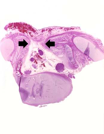
This is a low-power photomicrograph of one of the vertebral bodies from this case. In this section there are defects (arrows) in the vertebral body but the skin can be seen over the open vertebral canal.
File history
Click on a date/time to view the file as it appeared at that time.
| Date/Time | Thumbnail | Dimensions | User | Comment | |
|---|---|---|---|---|---|
| current | 05:45, 21 August 2013 | | 348 × 450 (16 KB) | Seung Park (talk | contribs) | This is a low-power photomicrograph of one of the vertebral bodies from this case. In this section there are defects (arrows) in the vertebral body but the skin can be seen over the open vertebral canal. |
- You cannot overwrite this file.
File usage
The following page links to this file: