File:IPLab13Myelomeningocele3.jpg
Revision as of 05:45, 21 August 2013 by Seung Park (talk | contribs) (This is a closer view of the previous gross photograph showing a normal lumbar vertebra from this case on the left. Once again, note the defect (arrow) in the vertebral body on the right due to failure of the vertebral column to close properly.)
No higher resolution available.
IPLab13Myelomeningocele3.jpg (746 × 450 pixels, file size: 17 KB, MIME type: image/jpeg)
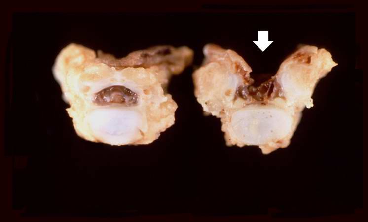
This is a closer view of the previous gross photograph showing a normal lumbar vertebra from this case on the left. Once again, note the defect (arrow) in the vertebral body on the right due to failure of the vertebral column to close properly.
File history
Click on a date/time to view the file as it appeared at that time.
| Date/Time | Thumbnail | Dimensions | User | Comment | |
|---|---|---|---|---|---|
| current | 05:45, 21 August 2013 | | 746 × 450 (17 KB) | Seung Park (talk | contribs) | This is a closer view of the previous gross photograph showing a normal lumbar vertebra from this case on the left. Once again, note the defect (arrow) in the vertebral body on the right due to failure of the vertebral column to close properly. |
- You cannot overwrite this file.
File usage
The following page links to this file: