File:IPLab13Myelomeningocele1.jpg
Revision as of 05:44, 21 August 2013 by Seung Park (talk | contribs) (This is a gross photograph of the fetus at autopsy. Note the defect in the lower lumbar region of the spinal column (arrow). The myelomeningocele can be seen protruding from this defect.)
No higher resolution available.
IPLab13Myelomeningocele1.jpg (697 × 450 pixels, file size: 27 KB, MIME type: image/jpeg)
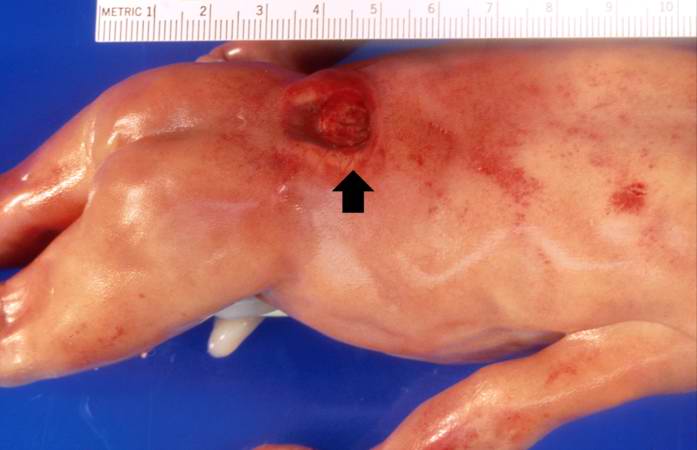
This is a gross photograph of the fetus at autopsy. Note the defect in the lower lumbar region of the spinal column (arrow). The myelomeningocele can be seen protruding from this defect.
A myelomeningocele is the herniation of the spinal cord--within the meninges--through a defect in the vertebral canal.
File history
Click on a date/time to view the file as it appeared at that time.
| Date/Time | Thumbnail | Dimensions | User | Comment | |
|---|---|---|---|---|---|
| current | 05:44, 21 August 2013 | | 697 × 450 (27 KB) | Seung Park (talk | contribs) | This is a gross photograph of the fetus at autopsy. Note the defect in the lower lumbar region of the spinal column (arrow). The myelomeningocele can be seen protruding from this defect. |
- You cannot overwrite this file.
File usage
The following page links to this file: