File:IPLab12COPD1.jpg
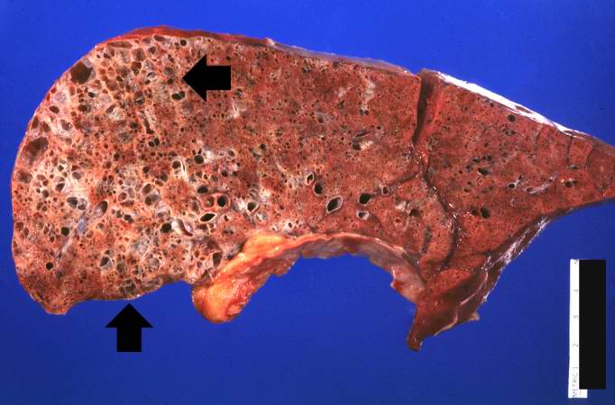
Revision as of 05:41, 21 August 2013 by Seung Park (talk | contribs) (This gross photograph of lung taken at autopsy demonstrates the degree of emphysematous change (arrows). Also note that the rest of the lung is consolidated, indicating a pneumonia.)
No higher resolution available.
IPLab12COPD1.jpg (682 × 450 pixels, file size: 45 KB, MIME type: image/jpeg)
This gross photograph of lung taken at autopsy demonstrates the degree of emphysematous change (arrows). Also note that the rest of the lung is consolidated, indicating a pneumonia.
Consolidation is the filling of lung air spaces with exudate--this is a sign of pneumonia.
In alcoholics, aspiration pneumonia is common--bacteria enter the lung via aspiration of gastric contents.
File history
Click on a date/time to view the file as it appeared at that time.
| Date/Time | Thumbnail | Dimensions | User | Comment | |
|---|---|---|---|---|---|
| current | 05:41, 21 August 2013 | | 682 × 450 (45 KB) | Seung Park (talk | contribs) | This gross photograph of lung taken at autopsy demonstrates the degree of emphysematous change (arrows). Also note that the rest of the lung is consolidated, indicating a pneumonia. |
- You cannot overwrite this file.
File usage
The following page links to this file: