File:IPLab12Mesothelioma2.jpg
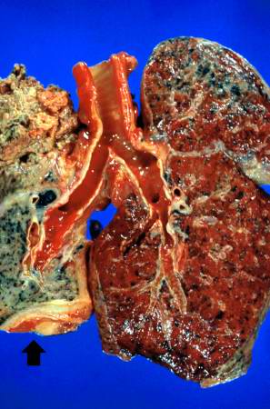
Revision as of 05:32, 21 August 2013 by Seung Park (talk | contribs) (This is a gross photograph of cut sections of the lungs. The right lung is congested. The left lung is shrunken and filled with tumor. There is a thick layer of tumor along the inferior portion of the lung (arrow).)
No higher resolution available.
IPLab12Mesothelioma2.jpg (297 × 450 pixels, file size: 30 KB, MIME type: image/jpeg)
This is a gross photograph of cut sections of the lungs. The right lung is congested. The left lung is shrunken and filled with tumor. There is a thick layer of tumor along the inferior portion of the lung (arrow).
File history
Click on a date/time to view the file as it appeared at that time.
| Date/Time | Thumbnail | Dimensions | User | Comment | |
|---|---|---|---|---|---|
| current | 05:32, 21 August 2013 | | 297 × 450 (30 KB) | Seung Park (talk | contribs) | This is a gross photograph of cut sections of the lungs. The right lung is congested. The left lung is shrunken and filled with tumor. There is a thick layer of tumor along the inferior portion of the lung (arrow). |
- You cannot overwrite this file.
File usage
The following page links to this file: