File:IPLab12RadiationChanges1.jpg
Revision as of 05:27, 21 August 2013 by Seung Park (talk | contribs) (This is a low-power photomicrograph of the surgical specimen of the ileum. The normal ileum is to the left (1). The area of stricture consists of dense fibrous connective tissue (2) and there is loss or marked atrophy of the epithelium (3).)
No higher resolution available.
IPLab12RadiationChanges1.jpg (675 × 450 pixels, file size: 26 KB, MIME type: image/jpeg)
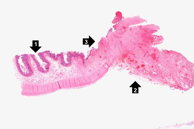
This is a low-power photomicrograph of the surgical specimen of the ileum. The normal ileum is to the left (1). The area of stricture consists of dense fibrous connective tissue (2) and there is loss or marked atrophy of the epithelium (3).
File history
Click on a date/time to view the file as it appeared at that time.
| Date/Time | Thumbnail | Dimensions | User | Comment | |
|---|---|---|---|---|---|
| current | 05:27, 21 August 2013 | | 675 × 450 (26 KB) | Seung Park (talk | contribs) | This is a low-power photomicrograph of the surgical specimen of the ileum. The normal ileum is to the left (1). The area of stricture consists of dense fibrous connective tissue (2) and there is loss or marked atrophy of the epithelium (3). |
- You cannot overwrite this file.
File usage
The following page links to this file: