File:IPLab12Alcoholic6.jpg
Revision as of 05:14, 21 August 2013 by Seung Park (talk | contribs)
No higher resolution available.
IPLab12Alcoholic6.jpg (607 × 450 pixels, file size: 42 KB, MIME type: image/jpeg)
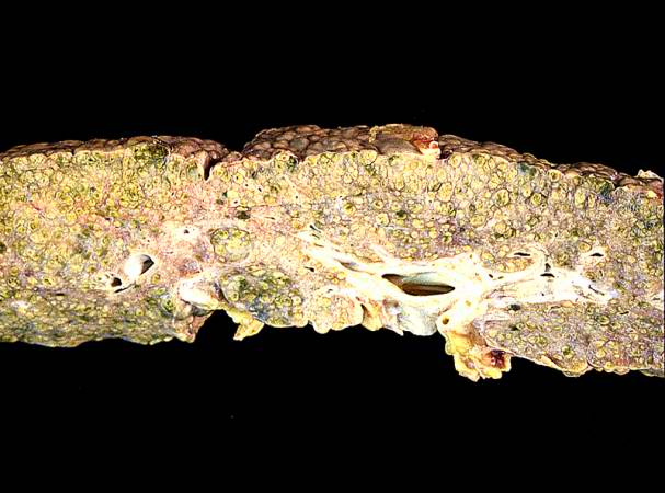
This photograph from another autopsy case shows another example of cirrhosis. Note the nodules, the fibrosis, the green coloration and the small size of this cirrhotic liver.
Cirrhosis is a liver disease characterized by necrosis, fibrosis, loss of normal liver architecture, and hyperplastic nodules.
File history
Click on a date/time to view the file as it appeared at that time.
| Date/Time | Thumbnail | Dimensions | User | Comment | |
|---|---|---|---|---|---|
| current | 05:13, 21 August 2013 | | 607 × 450 (42 KB) | Seung Park (talk | contribs) | In this high-power photomicrograph of trichrome-stained liver, the bands of fibrous tissue surround the hepatocyte nodules. There is some degeneration and dropout of hepatocytes in this nodule. Also note the increased numbers of bile ducts in the triad... |
- You cannot overwrite this file.
File usage
The following page links to this file: