File:IPLab11Cysticercosis3.jpg
No higher resolution available.
IPLab11Cysticercosis3.jpg (581 × 450 pixels, file size: 46 KB, MIME type: image/jpeg)
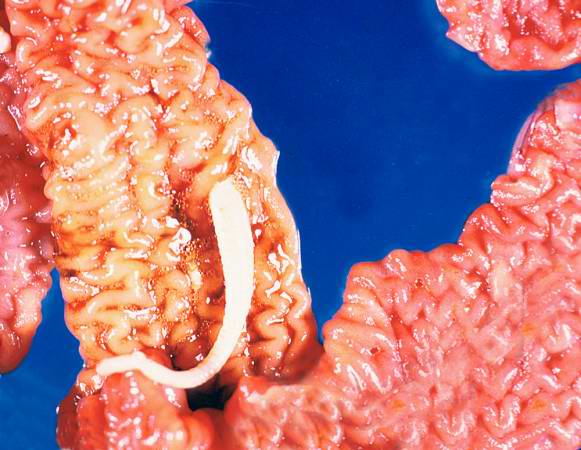
This photograph of an autopsy specimen from another patient shows an adult tapeworm in the intestine. Note that the worm attaches to the luminal surface of the intestine via the scolex.
File history
Click on a date/time to view the file as it appeared at that time.
| Date/Time | Thumbnail | Dimensions | User | Comment | |
|---|---|---|---|---|---|
| current | 00:04, 21 August 2013 | | 581 × 450 (46 KB) | Seung Park (talk | contribs) | This photograph of an autopsy specimen from another patient shows an adult tapeworm in the intestine. Note that the worm attaches to the luminal surface of the intestine via the scolex. |
You cannot overwrite this file.
File usage
The following page uses this file:
