IPLab:Lab 11:Leishmaniasis
Images[edit]
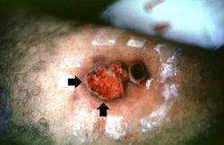
In this photograph of the skin lesion seen in this patient, note the raised edges (arrows) and the ulcerated center.
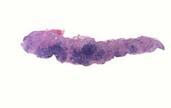
This is a low-power photomicrograph of the biopsy taken from this skin lesion. The ulcerated surface is at the top. Note that the specimen is heavily infiltrated with inflammatory cells.
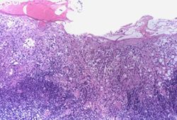
This is a higher-power photomicrograph of this biopsy. The ulcerated surface is seen on the top of the section. Again, note that the specimen is heavily infiltrated with inflammatory cells.
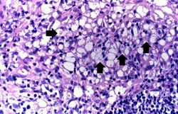
This high-power photomicrograph of the biopsy specimen shows more clearly the heavily infiltrate of inflammatory cells. Note the small blue structures inside the inflammatory cells (arrows).
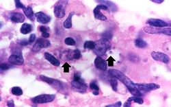
This is a high-power photomicrograph of an inflammatory cell containing cytoplasmic organisms (arrows).
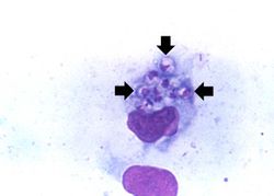
This is a high-power photomicrograph of a touch prep made from the skin lesion at the time of biopsy. A single macrophage can be seen with intracytoplasmic leishmania organisms (arrows).


| |||||
An infiltrate is an accumulation of cells in the lung parenchyma--this is a sign of pneumonia.