File list
This special page shows all uploaded files.
| Date | Name | Thumbnail | Size | User | Description | Versions |
|---|---|---|---|---|---|---|
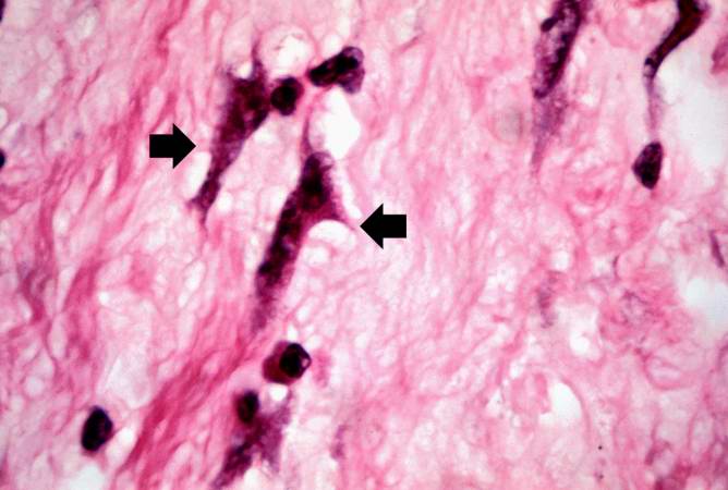
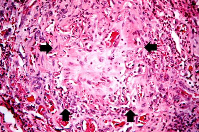
| 05:33, 21 August 2013 | IPLab12Mesothelioma6.jpg (file) |  |
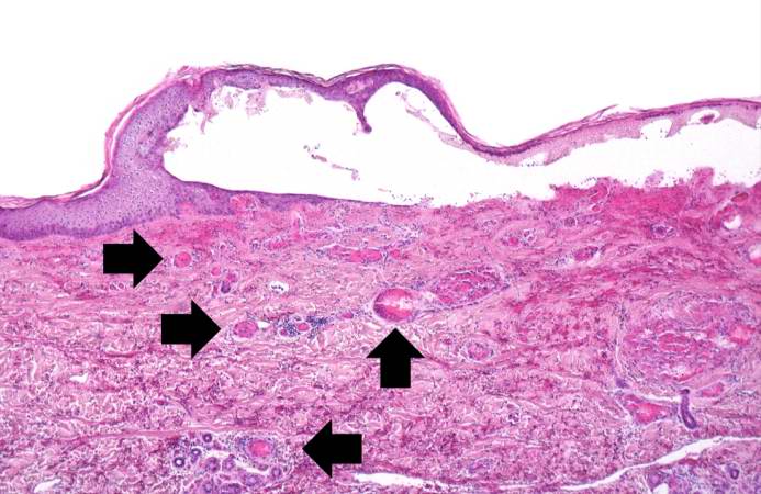
92 KB | Seung Park | In this higher-power photomicrograph the clusters of tumor cells (arrows) can be appreciated. | 1 |
| 05:33, 21 August 2013 | IPLab12Mesothelioma5.jpg (file) |  |
99 KB | Seung Park | This higher-power photomicrograph of left lung shows areas of consolidation and fibrosis (arrows). Also note that in many of these areas there are clusters of blue cells. | 1 |
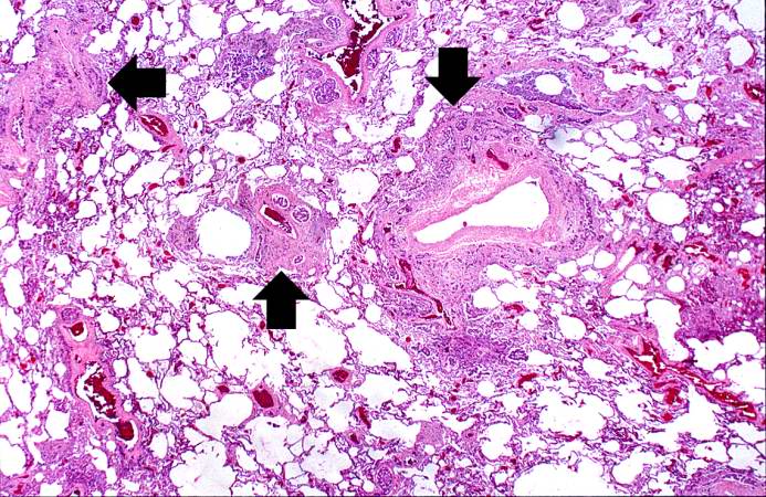
| 05:33, 21 August 2013 | IPLab12Mesothelioma4.jpg (file) |  |
113 KB | Seung Park | This is a low-power photomicrograph of a section of the left lung. At this magnification you can see areas of consolidation (arrows), especially around blood vessels. | 1 |
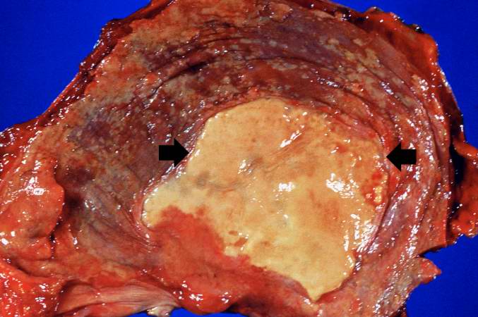
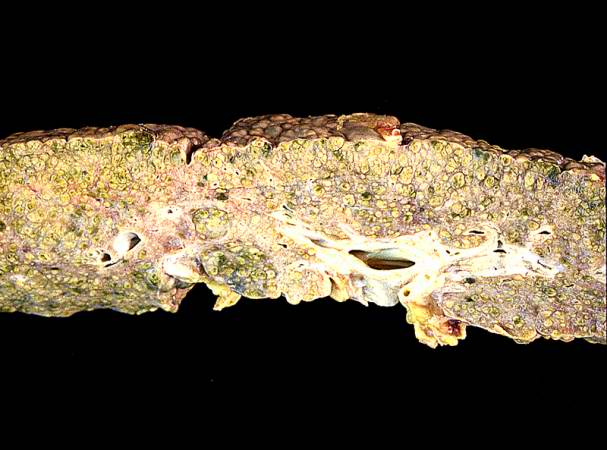
| 05:32, 21 August 2013 | IPLab12Mesothelioma3.jpg (file) |  |
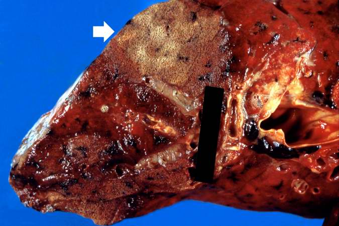
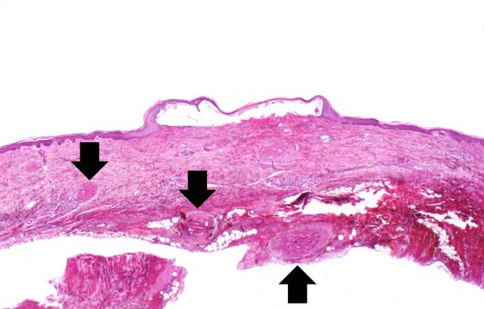
51 KB | Seung Park | This is a gross photograph of the thoracic surface of the diaphragm demonstrating the distinctive fibrocalcific plaque (arrows) produced by mesothelioma and multiple smaller plaques over the pleural surface of the diaphragm. | 1 |
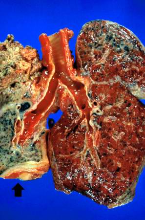
| 05:32, 21 August 2013 | IPLab12Mesothelioma2.jpg (file) |  |
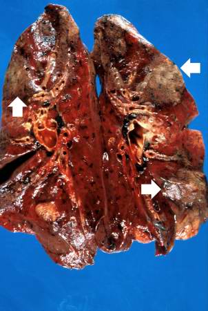
30 KB | Seung Park | This is a gross photograph of cut sections of the lungs. The right lung is congested. The left lung is shrunken and filled with tumor. There is a thick layer of tumor along the inferior portion of the lung (arrow). | 1 |
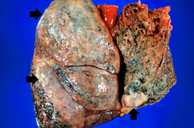
| 05:32, 21 August 2013 | IPLab12Mesothelioma1.jpg (file) |  |
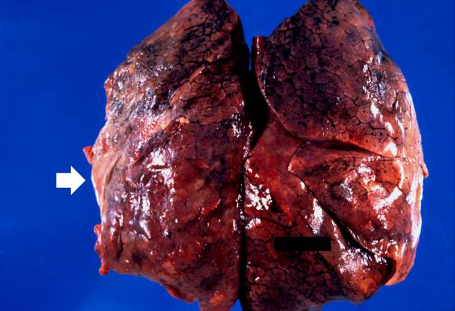
52 KB | Seung Park | This is a gross photograph of the lungs removed at autopsy. There is thickening of the pleural surface due to the tumor (arrows). The apical portion of the left lobe of the lung was ripped while trying to sever the adhesions between the lung and the ch... | 1 |
| 05:30, 21 August 2013 | IPLab12RadiationChanges8.jpg (file) |  |
38 KB | Seung Park | This high-power photomicrograph of the wall of the ileum shows more examples of pleomorphic cells caused by radiation injury (arrows). | 1 |
| 05:29, 21 August 2013 | IPLab12RadiationChanges7.jpg (file) |  |
47 KB | Seung Park | This high-power photomicrograph of the wall of the ileum shows more examples of pleomorphic cells (arrows) due to radiation injury. | 1 |
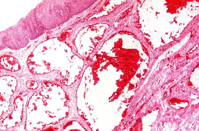
| 05:29, 21 August 2013 | IPLab12RadiationChanges5.jpg (file) |  |
81 KB | Seung Park | This is a high-power photomicrograph of the wall of the ileum showing a blood vessel that has suffered radiation-induced damage and is completely occluded (arrows). | 1 |
| 05:28, 21 August 2013 | IPLab12RadiationChanges6.jpg (file) |  |
57 KB | Seung Park | This is a high-power photomicrograph of the wall of the ileum showing a blood vessel that has suffered radiation-induced damage and is completely occluded (arrows). | 1 |
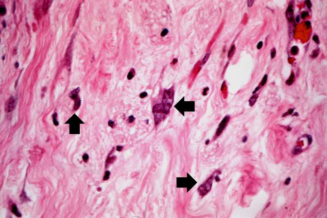
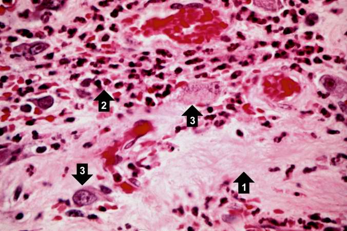
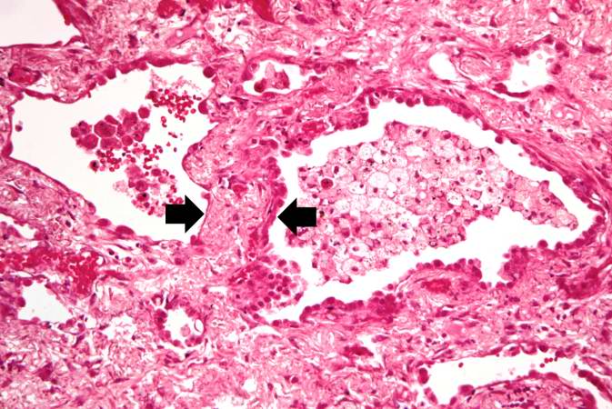
| 05:28, 21 August 2013 | IPLab12RadiationChanges4.jpg (file) |  |
39 KB | Seung Park | This is a high-power photomicrograph showing the atrophied epithelium in the area of radiation injury (1). Note the dense fibrous connective tissue within the wall of the ileum and the congested blood vessels (2). | 1 |
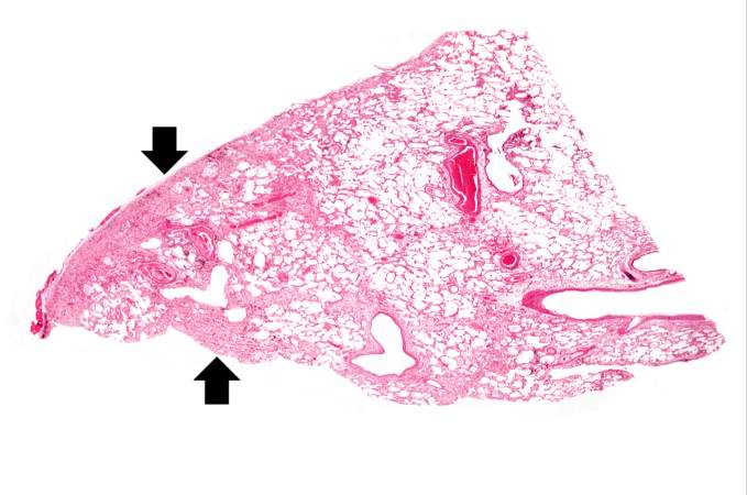
| 05:28, 21 August 2013 | IPLab12RadiationChanges3.jpg (file) |  |
38 KB | Seung Park | This is a higher-power photomicrograph showing the atrophied epithelium in the area of radiation injury. There are some epithelial cells deep within the mucosa (1). Note the dense fibrous connective tissue (2) within the wall of the ileum. | 1 |
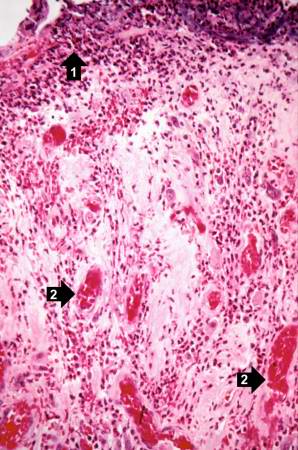
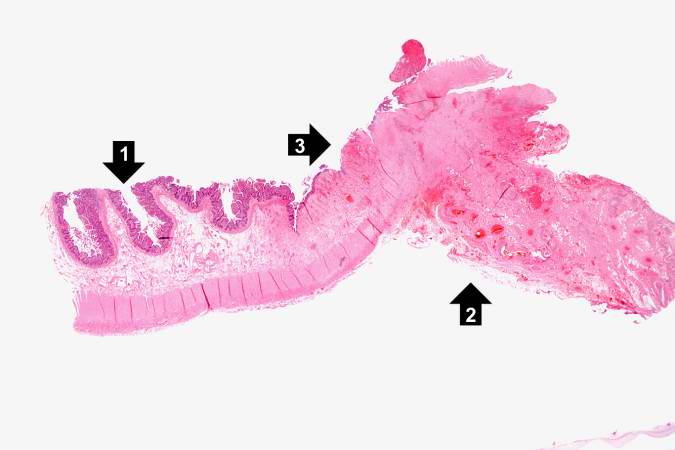
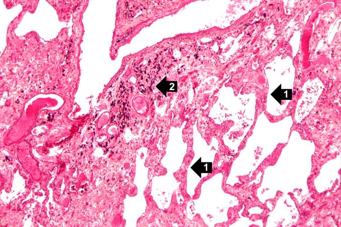
| 05:28, 21 August 2013 | IPLab12RadiationChanges2.jpg (file) |  |
26 KB | Seung Park | This is a higher-power photomicrograph of the surgical specimen of the ileum showing the transition from the normal epithelium (1) to the atrophied epithelium (2) in the area of radiation injury. | 1 |
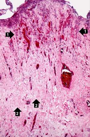
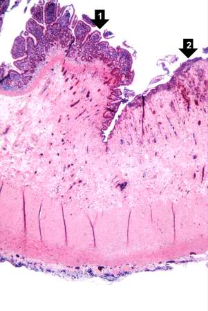
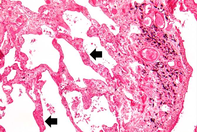
| 05:27, 21 August 2013 | IPLab12RadiationChanges1.jpg (file) |  |
26 KB | Seung Park | This is a low-power photomicrograph of the surgical specimen of the ileum. The normal ileum is to the left (1). The area of stricture consists of dense fibrous connective tissue (2) and there is loss or marked atrophy of the epithelium (3). | 1 |
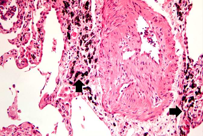
| 05:25, 21 August 2013 | IPLab12RadiationFibrosis13.jpg (file) |  |
69 KB | Seung Park | This is a high-power photomicrograph of a recanalized blood vessel in the lung. Notice the anthracotic pigment adjacent to the vessel (arrows). | 1 |
| 05:25, 21 August 2013 | IPLab12RadiationFibrosis12.jpg (file) |  |
72 KB | Seung Park | This high-power photomicrograph shows intimal changes (arrows) in this blood vessel in the lung. | 1 |
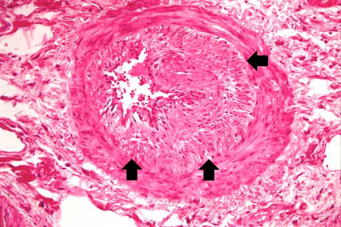
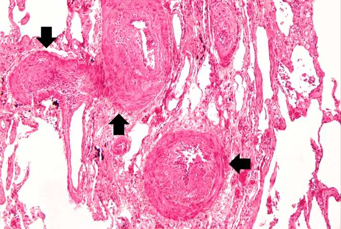
| 05:25, 21 August 2013 | IPLab12RadiationFibrosis11.jpg (file) |  |
73 KB | Seung Park | This medium-power photomicrograph shows fibrosis and severe intimal changes in blood vessels (arrows). | 1 |
| 05:25, 21 August 2013 | IPLab12RadiationFibrosis10.jpg (file) |  |
80 KB | Seung Park | This is another high-power photomicrograph of an area of tissue with diffuse fibrosis and thickening of the alveolar septa. There are also accumulations of anthracotic pigment in this area (arrows). | 1 |
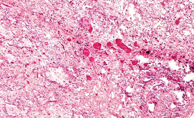
| 05:25, 21 August 2013 | IPLab12RadiationFibrosis9.jpg (file) |  |
107 KB | Seung Park | This is a photomicrograph of an area of tissue exhibiting diffuse fibrosis and thickening of the alveolar septa. | 1 |
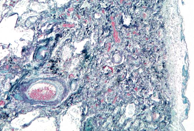
| 05:24, 21 August 2013 | IPLab12RadiationFibrosis8.jpg (file) |  |
75 KB | Seung Park | This is a photomicrograph of a trichrome-stained section of lung demonstrating the extensive fibrosis throughout this section (green-blue stained material is fibrous connective tissue). | 1 |
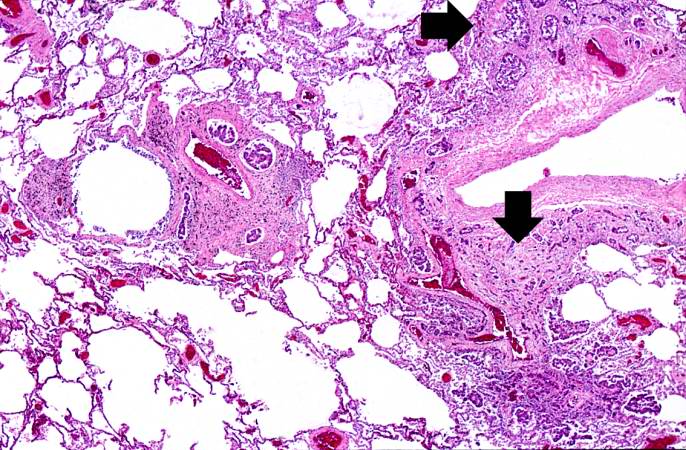
| 05:24, 21 August 2013 | IPLab12RadiationFibrosis7.jpg (file) |  |
75 KB | Seung Park | This high-power photomicrograph of lung section shows the thickening of the alveolar septum (arrows) by fibrous connective tissue. | 1 |
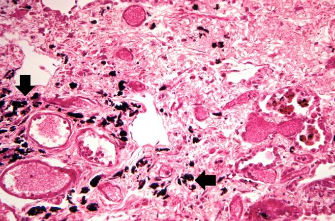
| 05:24, 21 August 2013 | IPLab12RadiationFibrosis6.jpg (file) |  |
71 KB | Seung Park | This is another high-power photomicrograph of lung section showing the thickening of the alveolar septa (arrows) and accumulations of black anthracotic pigment. | 1 |
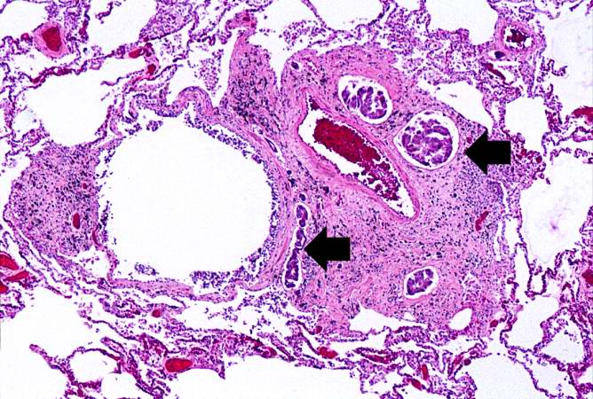
| 05:24, 21 August 2013 | IPLab12RadiationFibrosis5.jpg (file) |  |
76 KB | Seung Park | This is a higher-power photomicrograph of lung section. Note the thickening of the alveolar septa (1) and accumulations of anthracotic pigment (2). | 1 |
| 05:23, 21 August 2013 | IPLab12RadiationFibrosis4.jpg (file) |  |
41 KB | Seung Park | This is a low-power photomicrograph of lung section. Note the thickening of the alveolar septa (arrows). | 1 |
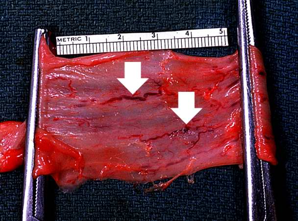
| 05:23, 21 August 2013 | IPLab12RadiationFibrosis3.jpg (file) |  |
53 KB | Seung Park | This is a gross photograph showing a closer view of a cut section of lung. An area of fibrosis (arrow) is evident in this photograph. | 1 |
| 05:23, 21 August 2013 | IPLab12RadiationFibrosis2.jpg (file) |  |
27 KB | Seung Park | This is a gross photograph of cut sections of lung. There are several areas of fibrosis (arrows) within the lung parenchyma. | 1 |
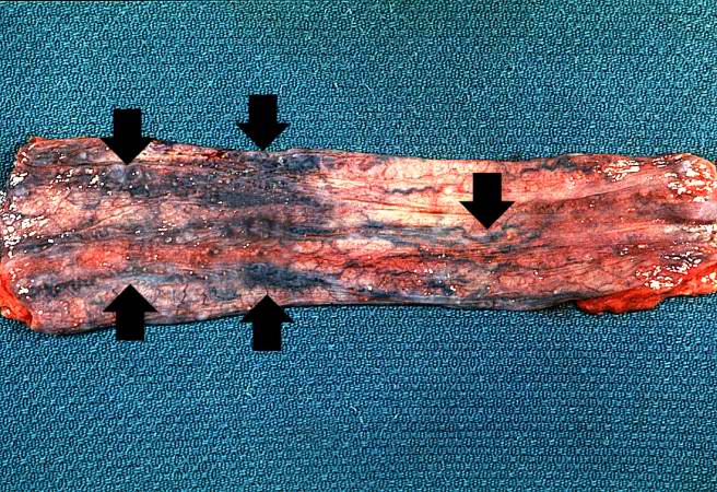
| 05:23, 21 August 2013 | IPLab12RadiationFibrosis1.jpg (file) |  |
43 KB | Seung Park | This is a gross photograph of lung demonstrating areas of fibrosis on the pleural surface (arrow). | 1 |
| 05:20, 21 August 2013 | IPLab12Acetaminophen8.jpg (file) |  |
59 KB | Seung Park | This is a higher-power photomicrograph of the skin from this patient showing a blister and the thrombosed small vessels (arrows) in the dermis. | 1 |
| 05:20, 21 August 2013 | IPLab12Acetaminophen7.jpg (file) |  |
40 KB | Seung Park | This low-power photomicrograph of the skin from this patient shows a blister and numerous thrombosed vessels (arrows) in the dermis. | 1 |
| 05:20, 21 August 2013 | IPLab12Acetaminophen6.jpg (file) |  |
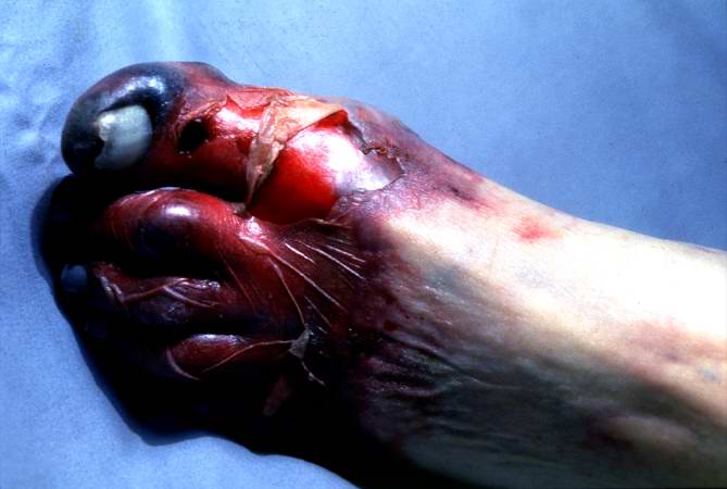
32 KB | Seung Park | Photograph taken at autopsy demonstrating the severe necrosis of the skin of foot. | 1 |
| 05:20, 21 August 2013 | IPLab12Acetaminophen5.jpg (file) |  |
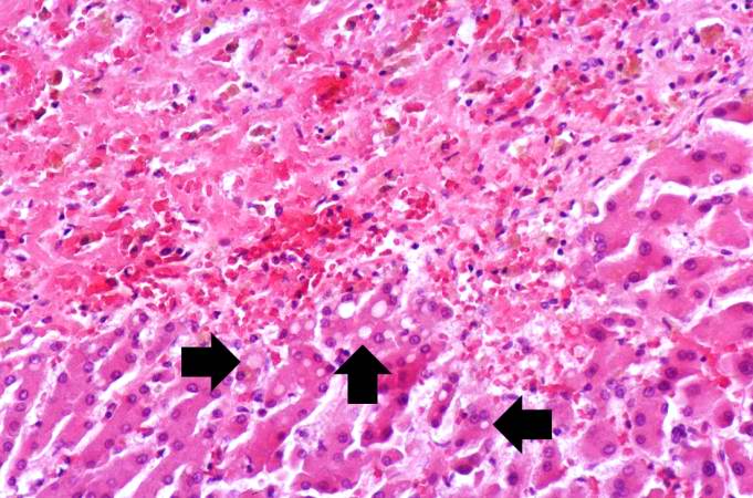
71 KB | Seung Park | This is a high-power photomicrograph of the edge of the necrosis. Again, note the coagulation necrosis and hemorrhage in this area. The viable hepatocytes at the edge of this necrosis are vacuolated (arrows). | 1 |
| 05:19, 21 August 2013 | IPLab12Acetaminophen4.jpg (file) |  |
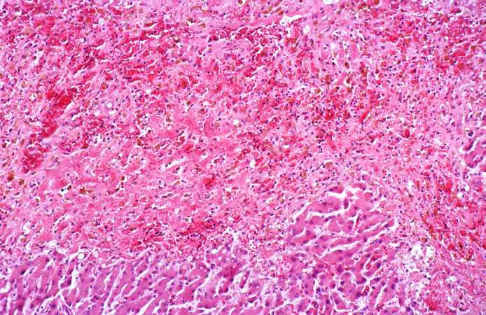
95 KB | Seung Park | This is a medium-power photomicrograph of the areas of hemorrhagic necrosis. Note the coagulation necrosis and hemorrhage in this area. Viable hepatocytes can be seen along the edge of this lesion. | 1 |
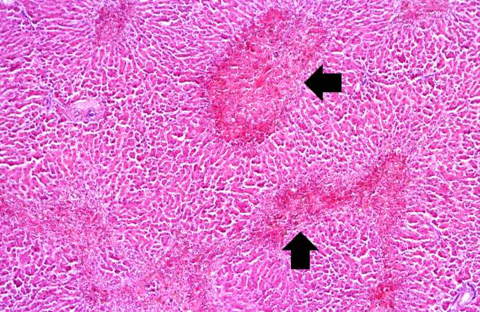
| 05:19, 21 August 2013 | IPLab12Acetaminophen3.jpg (file) |  |
100 KB | Seung Park | This is another example of the areas of hemorrhagic necrosis (arrows). | 1 |
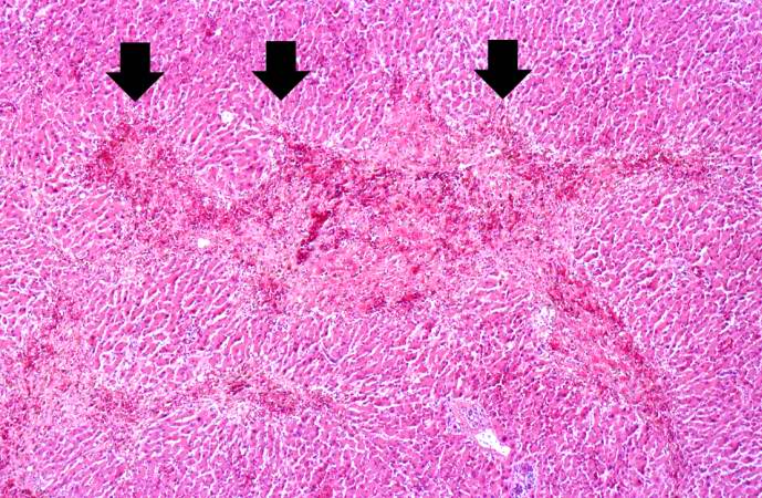
| 05:19, 21 August 2013 | IPLab12Acetaminophen2.jpg (file) |  |
103 KB | Seung Park | In this photomicrograph of the liver from this patient there are areas of hemorrhagic necrosis (arrows). | 1 |
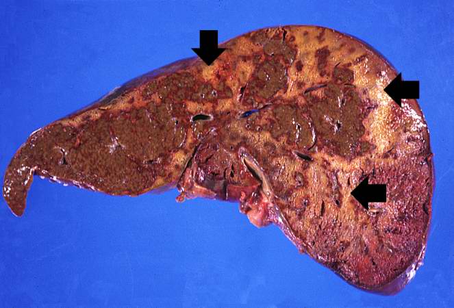
| 05:19, 21 August 2013 | IPLab12Acetaminophen1.jpg (file) |  |
48 KB | Seung Park | This gross photograph of the liver from this patient demonstrates the pale areas of necrosis (arrows). | 1 |
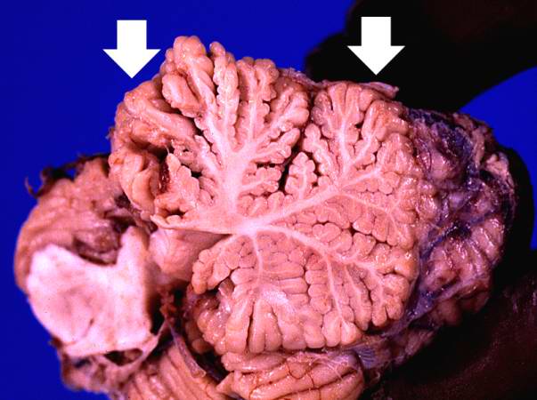
| 05:17, 21 August 2013 | IPLab12Alcoholic14.jpg (file) |  |
30 KB | Seung Park | This is another photograph of the cerebellum from this patient demonstrating the marked thinning of the anterior portion of the cerebellum (arrows). This pattern of cerebellar damage is consistent with Wernicke's encephalopathy. | 1 |
| 05:16, 21 August 2013 | IPLab12Alcoholic13.jpg (file) |  |
41 KB | Seung Park | This photograph of the cerebellum from this patient demonstrates the marked thinning of the anterior portion of the cerebellum (arrows). | 1 |
| 05:16, 21 August 2013 | IPLab12Alcoholic12.jpg (file) |  |
82 KB | Seung Park | This photomicrograph shows the dilated vessel just under the epithelium of the esophagus. | 1 |
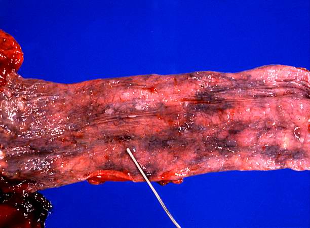
| 05:16, 21 August 2013 | IPLab12Alcoholic11.jpg (file) |  |
58 KB | Seung Park | This photograph taken from still another patient at autopsy demonstrates the esophageal varices in the distal esophagus (arrows). The esophagus was clamped before removing the esophagus from the body in order to trap the blood in these distended varice... | 1 |
| 05:16, 21 August 2013 | IPLab12Alcoholic10.jpg (file) |  |
105 KB | Seung Park | This is a photograph taken from another patient at autopsy to demonstrate numerous esophageal varices in the distal esophagus (arrows). None of these varices have ruptured. | 1 |
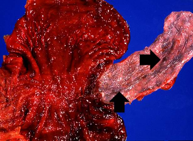
| 05:15, 21 August 2013 | IPLab12Alcoholic9.jpg (file) |  |
41 KB | Seung Park | In this closer view of the distal esophagus the ruptured varix is indicated by the probe. Other varices and areas of submucosal hemorrhage can also be appreciated. | 1 |
| 05:15, 21 August 2013 | IPLab12Alcoholic8.jpg (file) |  |
57 KB | Seung Park | This photograph taken at autopsy shows the distal portion of the esophagus and the stomach. The esophageal varices are visible just under the mucosa of the esophagus (arrows). Some of the blood from the ruptured varix can still be seen in the stomach. | 1 |
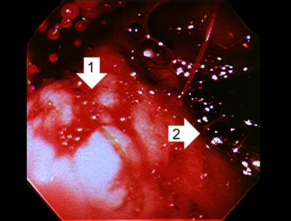
| 05:15, 21 August 2013 | IPLab12Alcoholic7.jpg (file) |  |
32 KB | Seung Park | This photograph was taken during the EGD while the patient was alive. Note the red hyperemic areas (1) and the area of hemorrhage (2). | 1 |
| 05:14, 21 August 2013 | IPLab12Alcoholic5.jpg (file) |  |
122 KB | Seung Park | In this high-power photomicrograph of trichrome-stained liver, the bands of fibrous tissue surround the hepatocyte nodules. There is some degeneration and dropout of hepatocytes in this nodule. Also note the increased numbers of bile ducts in the triad... | 1 |
| 05:13, 21 August 2013 | IPLab12Alcoholic6.jpg (file) |  |
42 KB | Seung Park | In this high-power photomicrograph of trichrome-stained liver, the bands of fibrous tissue surround the hepatocyte nodules. There is some degeneration and dropout of hepatocytes in this nodule. Also note the increased numbers of bile ducts in the triad... | 1 |
| 05:13, 21 August 2013 | IPLab12Alcoholic4.jpg (file) |  |
130 KB | Seung Park | In this is medium-power photomicrograph of trichrome stained liver the bands of fibrous tissue are seen to form "bridges" between triad areas (arrows); this is called "bridging fibrosis." Also note the fibrous tissue (arrows) and how the hepatocytes ar... | 1 |
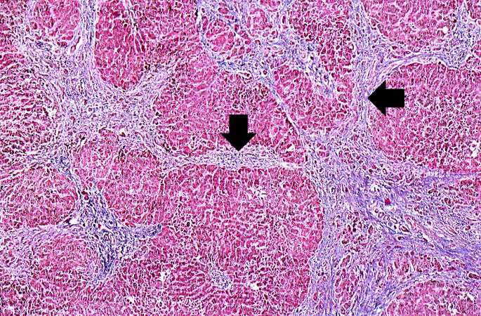
| 05:13, 21 August 2013 | IPLab12Alcoholic3.jpg (file) |  |
102 KB | Seung Park | This is a low-power photomicrograph of this liver stained with a trichrome stain to highlight the fibrous tissue (arrows). Also note the nodular pattern. | 1 |
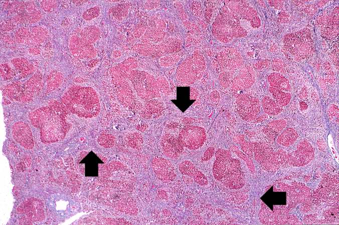
| 05:13, 21 August 2013 | IPLab12Alcoholic2.jpg (file) |  |
65 KB | Seung Park | This is a closer view of the liver from this patient. Again note the nodular pattern and the areas of greenish discoloration. These green nodules are actually the viable hepatocytes that are stained green because of bile stasis. The pale areas are the ... | 1 |
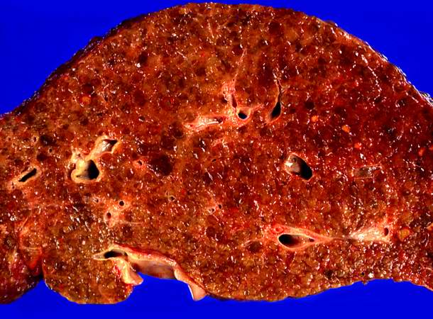
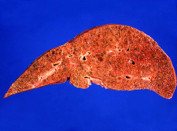
| 05:13, 21 August 2013 | IPLab12Alcoholic1.jpg (file) |  |
40 KB | Seung Park | This is a gross photograph of the liver from this patient. Note the nodular pattern and the areas of greenish discoloration as well as pale tan areas. | 1 |
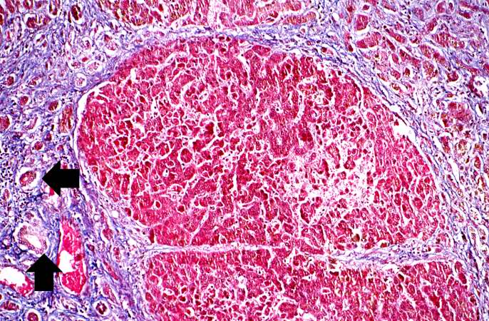
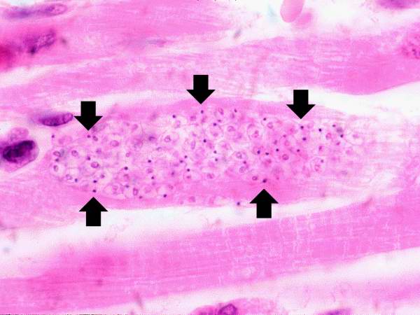
| 05:08, 21 August 2013 | IPLab11Chagas6.jpg (file) |  |
34 KB | Seung Park | This is a higher-power photomicrograph of an H & E stained heart biopsy from this patient. Note the T. cruzi amastigotes (arrows) within this longitudinal section of a myocyte. | 1 |