File list
This special page shows all uploaded files.
| Date | Name | Thumbnail | Size | User | Description | Versions |
|---|---|---|---|---|---|---|
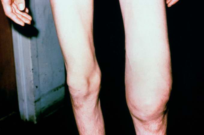
| 02:13, 21 August 2013 | IPLab7Osteosarcoma1.jpg (file) |  |
19 KB | Seung Park | This is a photograph of the patient prior to surgery. Note the marked swelling of the knee. | 1 |
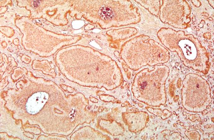
| 02:08, 21 August 2013 | IPLab7Carcinoid11.jpg (file) |  |
70 KB | Seung Park | This is a high-power view of the same section stained with a silver stain to delineate carcinoid tumor cells (brown). | 1 |
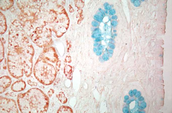
| 02:08, 21 August 2013 | IPLab7Carcinoid10.jpg (file) |  |
45 KB | Seung Park | This is a higher-power view of the previous section stained with a silver stain to delineate carcinoid tumor cells (brown) and a mucin stain (blue) to stain the glands. | 1 |
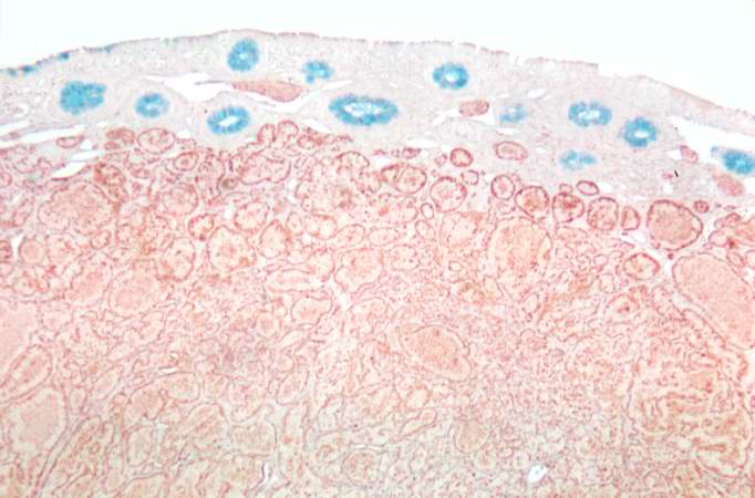
| 02:08, 21 August 2013 | IPLab7Carcinoid9.jpg (file) |  |
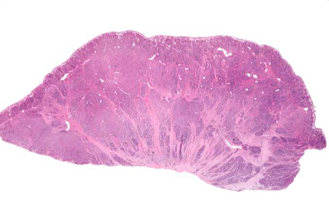
46 KB | Seung Park | This is a low-power photomicrograph of a section of cecum containing tumor stained to demonstrate the secretory granules in these tumor cells (brown-colored stain). The blue color is the mucin in the glands just under the mucosal surface. | 1 |
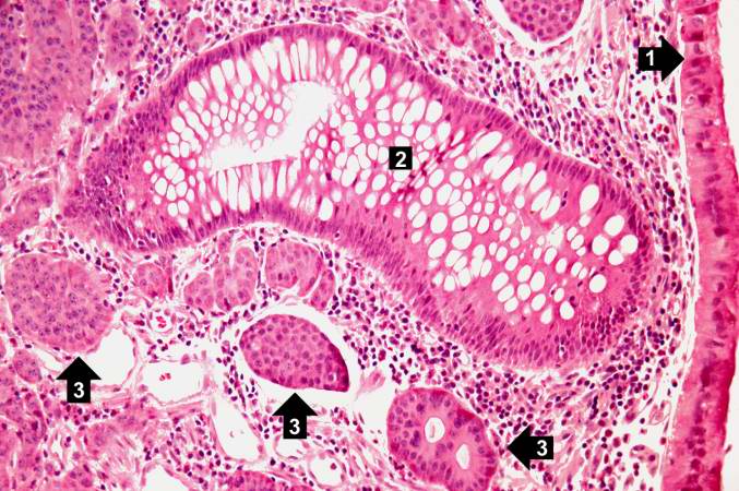
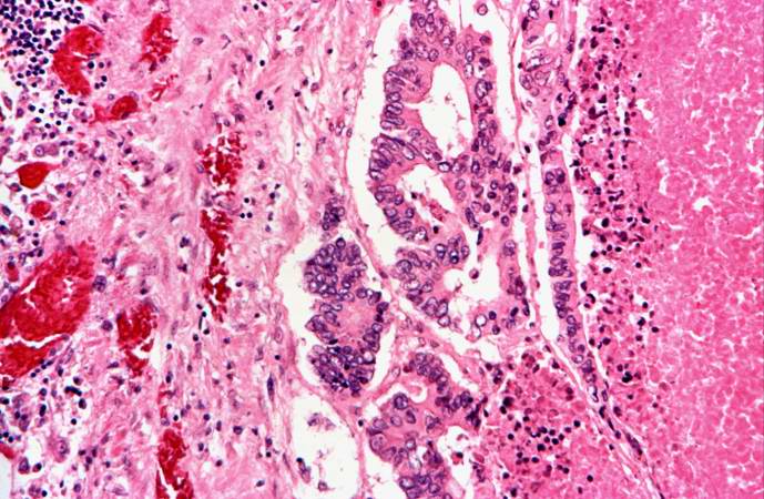
| 02:08, 21 August 2013 | IPLab7Carcinoid8.jpg (file) |  |
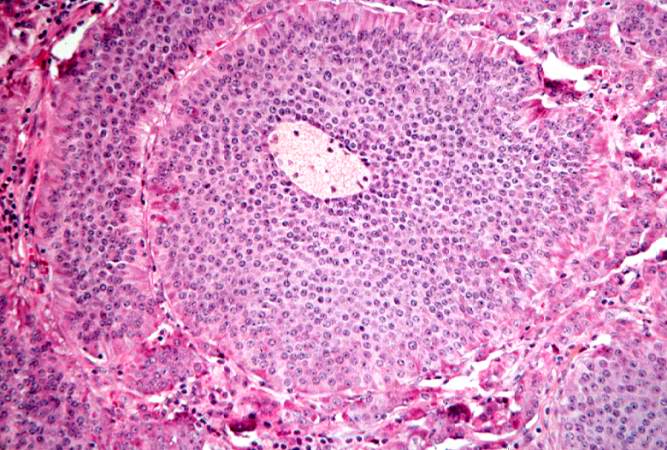
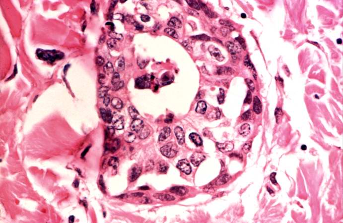
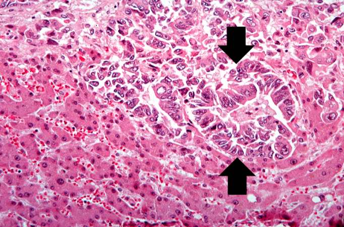
81 KB | Seung Park | This is a higher-power photomicrograph of the previous section showing intact mucosa (1), a gland (2), and the submucosal carcinoid tumor cells (3). | 1 |
| 02:07, 21 August 2013 | IPLab7Carcinoid7.jpg (file) |  |
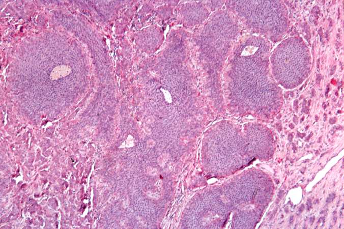
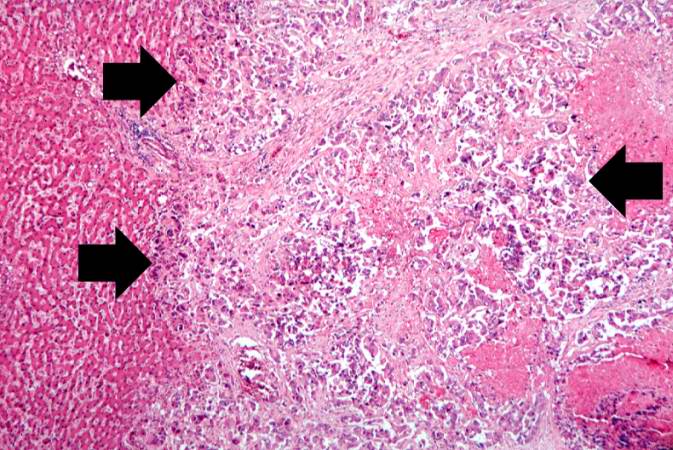
70 KB | Seung Park | This is a low-power photomicrograph of another one of the subcutaneous masses in the cecum. The mucosa is normal and the tumor cells are in the submucosa. | 1 |
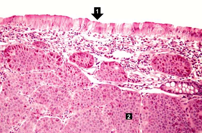
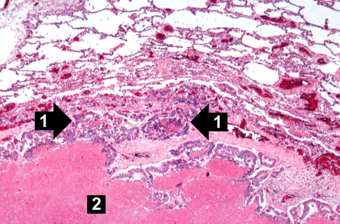
| 02:07, 21 August 2013 | IPLab7Carcinoid6.jpg (file) |  |
59 KB | Seung Park | This is a higher-power photomicrograph of the previous section showing the intact mucosa (right) and the submucosal carcinoid tumor. | 1 |
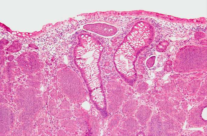
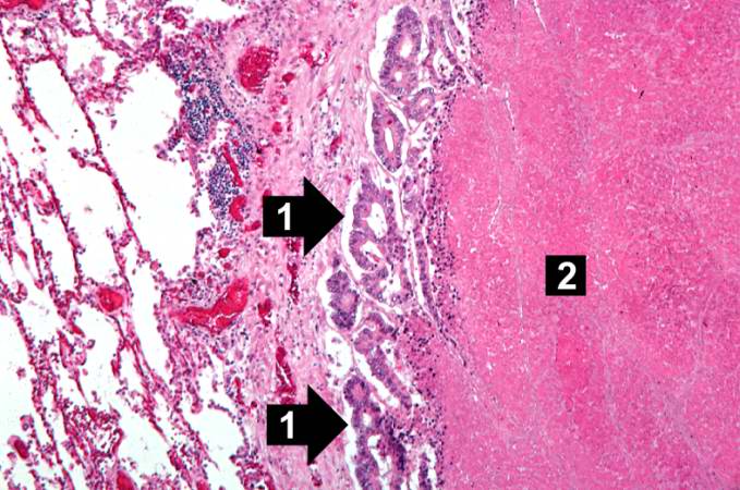
| 02:07, 21 August 2013 | IPLab7Carcinoid5.jpg (file) |  |
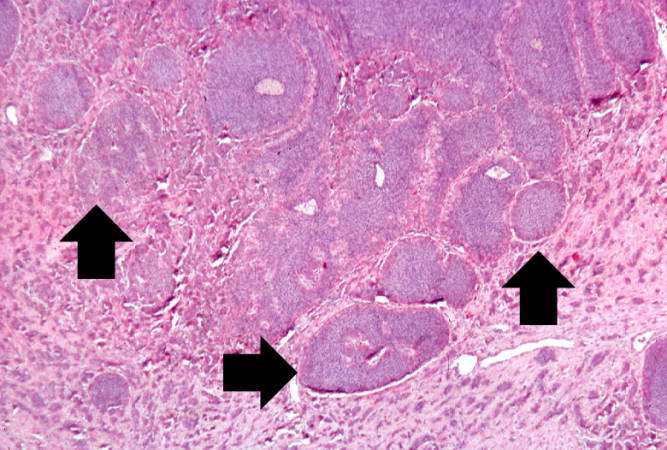
65 KB | Seung Park | This is a low-power photomicrograph of one of the subcutaneous masses in the cecum. Note that the mucosa (1) is virtually normal and the tumor cells are in the submucosa (2). | 1 |
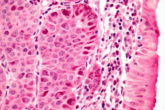
| 02:07, 21 August 2013 | IPLab7Carcinoid4.jpg (file) |  |
77 KB | Seung Park | This is a high-power photomicrograph of the surgical specimen showing the cellular morphology. The tumor cells are monotonously similar with scant, pink, granular cytoplasm and a round-to-oval stippled nucleus. As in most carcinoid tumors, there is min... | 1 |
| 02:06, 21 August 2013 | IPLab7Carcinoid3.jpg (file) |  |
72 KB | Seung Park | This is a high-power photomicrograph of the surgical specimen showing the tumor's growth pattern--cells form discrete islands, trabeculae, and glands. | 1 |
| 02:06, 21 August 2013 | IPLab7Carcinoid2.jpg (file) |  |
62 KB | Seung Park | This is a higher-power photomicrograph of the surgical specimen showing nests of tumor cells (arrows). | 1 |
| 02:06, 21 August 2013 | IPLab7Carcinoid1.jpg (file) |  |
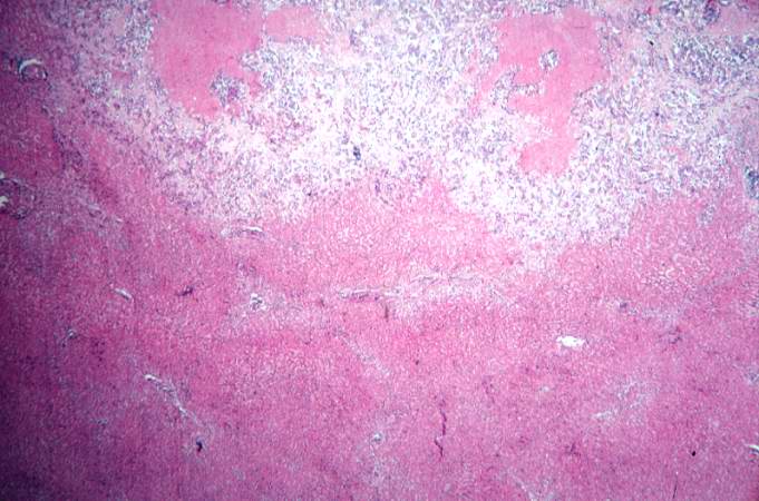
31 KB | Seung Park | This is a low-power photomicrograph of the surgical specimen showing basophilic and eosinophilic areas delimiting areas of tumor infiltration. | 1 |
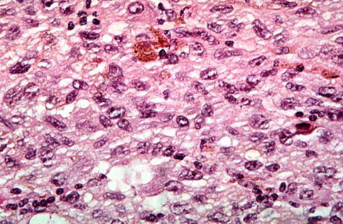
| 02:01, 21 August 2013 | IPLab7Bronchogenic9.jpg (file) |  |
73 KB | Seung Park | This high-power photomicrograph of tumor shows the cytologic detail of a less-differentiated area of neoplasm with cellular anaplasia. | 1 |
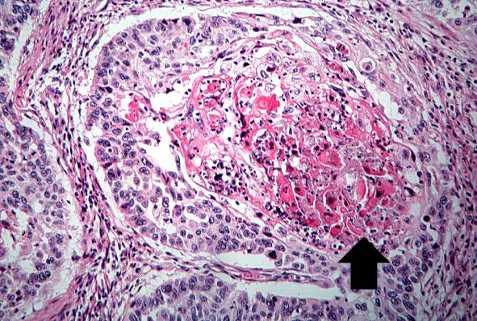
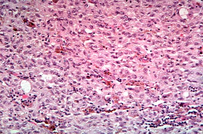
| 02:01, 21 August 2013 | IPLab7Bronchogenic8.jpg (file) |  |
91 KB | Seung Park | This is a high power photomicrograph of tumor with an area of central necrosis (arrow). | 1 |
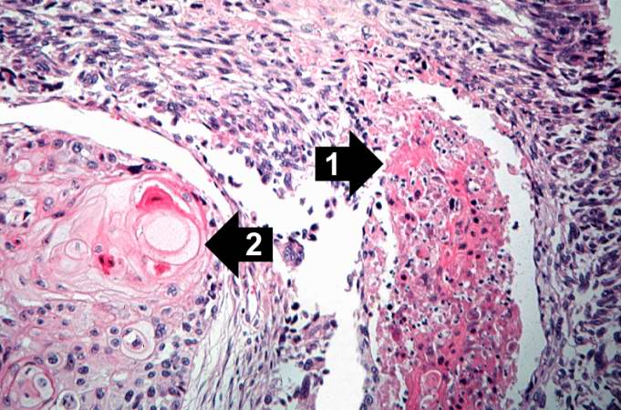
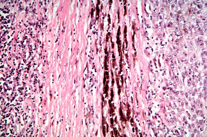
| 02:01, 21 August 2013 | IPLab7Bronchogenic7.jpg (file) |  |
75 KB | Seung Park | This is a high-power photomicrograph showing cytologic detail of the tumor with an area of necrosis (1) and a more differentiated area with keratin pearl formation (2). | 1 |
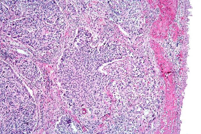
| 02:00, 21 August 2013 | IPLab7Bronchogenic6.jpg (file) |  |
98 KB | Seung Park | This is a photomicrograph of tumor from an area of invasion with compression of fibrous stroma and focal necrosis. | 1 |
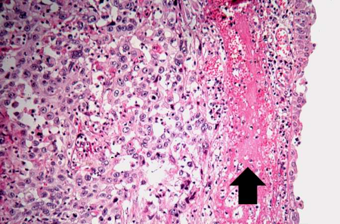
| 02:00, 21 August 2013 | IPLab7Bronchogenic5.jpg (file) |  |
71 KB | Seung Park | This is a higher-power photomicrograph of the mucosal surface (right) with an area of hemorrhage (arrow) and underlying tumor (left). | 1 |
| 02:00, 21 August 2013 | IPLab7Bronchogenic4.jpg (file) |  |
99 KB | Seung Park | This is a higher-power photomicrograph of bronchus with the ulcerated mucosal surface on the right and tumor underneath. | 1 |
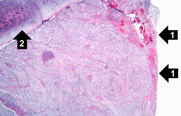
| 02:00, 21 August 2013 | IPLab7Bronchogenic3.jpg (file) |  |
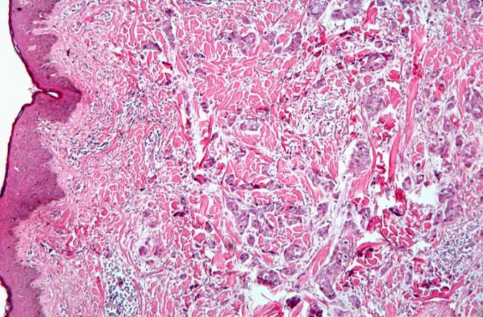
58 KB | Seung Park | This is a photomicrograph of bronchus with ulcerated mucosal surface on the right (1). The submucosa is completely filled with tumor down to the cartilage (2). | 1 |
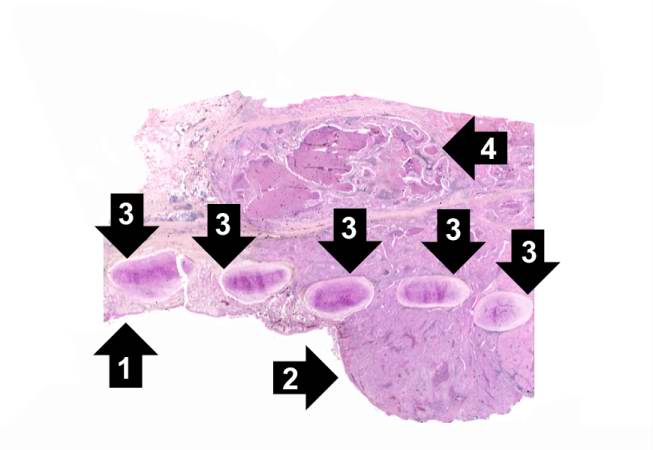
| 02:00, 21 August 2013 | IPLab7Bronchogenic2.jpg (file) |  |
25 KB | Seung Park | This is a low-power photomicrograph of bronchus showing normal mucosa (1) with transition to carcinoma (2). Note the bronchial cartilage (3) and the invasion of tumor through the entire wall of the bronchus with tumor extending to the serosal surface (4). | 1 |
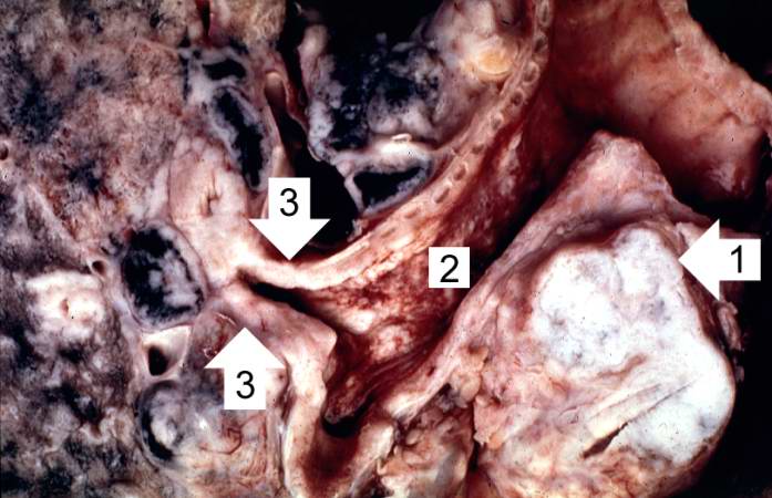
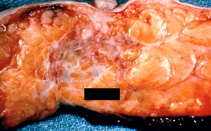
| 01:59, 21 August 2013 | IPLab7Bronchogenic1.jpg (file) |  |
53 KB | Seung Park | This is a gross photograph of bronchogenic carcinoma. The large tumor mass can be seen adjacent to the bronchus (1). Note that the epithelial surface of the bronchus is rough and irregular (2). The first branch off the right main stem bronchus is parti... | 1 |
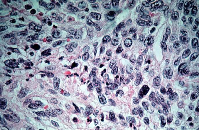
| 01:57, 21 August 2013 | IPLab7Melanoma8.jpg (file) |  |
76 KB | Seung Park | This is a high-power photomicrograph of the main tumor mass showing the cellular details. The individual melanoma cells contain large nuclei with irregular contours having chromatin clumped at the periphery of the nuclear membrane and prominent red (eo... | 1 |
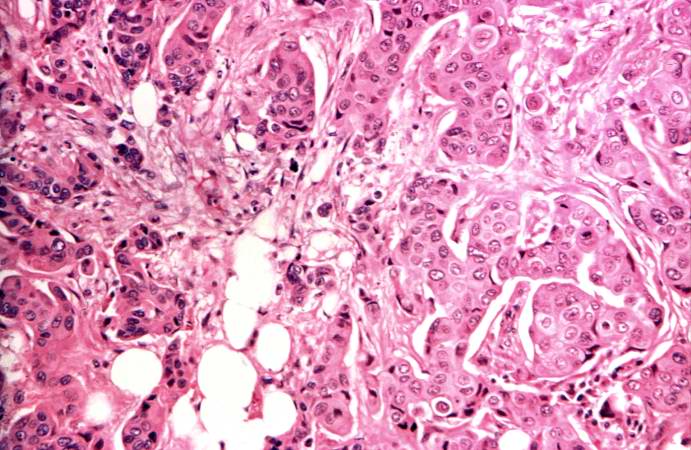
| 01:56, 21 August 2013 | IPLab7Melanoma7.jpg (file) |  |
81 KB | Seung Park | This is a high-power photomicrograph of the main tumor mass with the cells growing as poorly formed nests and sheets of cells. There is little if any pigment in this section. | 1 |
| 01:56, 21 August 2013 | IPLab7Melanoma6.jpg (file) |  |
82 KB | Seung Park | This is a higher magnification showing the abundant extracellular melanin surrounding the tumor cells (brown pigment). | 1 |
| 01:56, 21 August 2013 | IPLab7Melanoma5.jpg (file) |  |
93 KB | Seung Park | This is a higher-magnification showing the abundant extracellular melanin (arrows) surrounding the tumor cells. This section of neoplasm shows the numerous cells with abundant cytoplasm and brown pigment within the cytoplasm of some of these cells. | 1 |
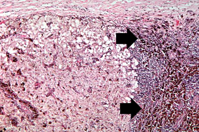
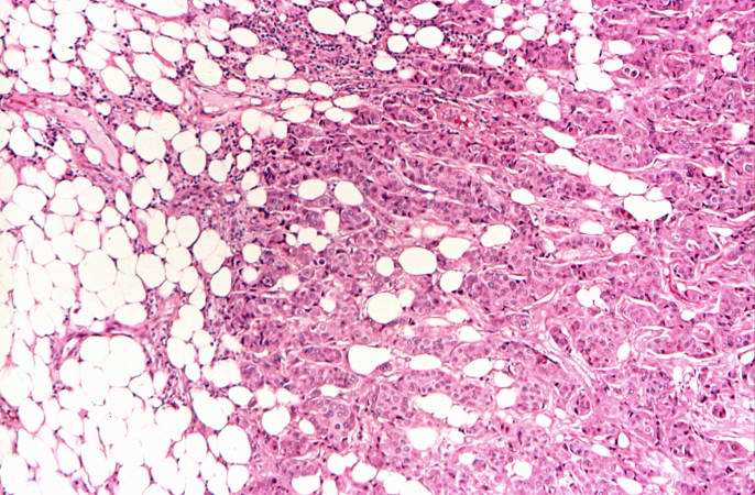
| 01:56, 21 August 2013 | IPLab7Melanoma4.jpg (file) |  |
74 KB | Seung Park | This higher-power photomicrograph shows the remaining portion of lymph node (arrow). The rest of the lymph node is invaded by a neoplasm composed of cells with lighter eosinophilic cytoplasm and pigment. | 1 |
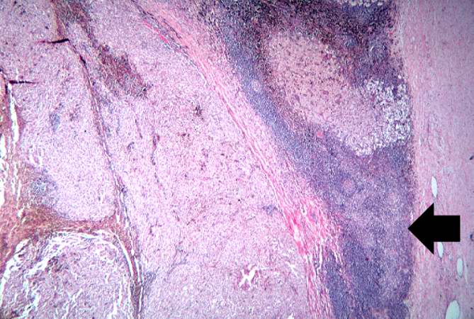
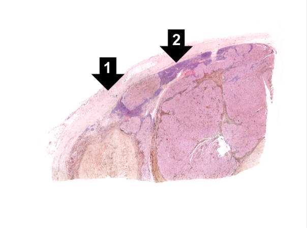
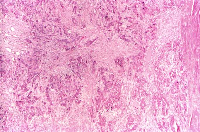
| 01:55, 21 August 2013 | IPLab7Melanoma3.jpg (file) |  |
21 KB | Seung Park | This is a low-power photomicrograph of lymph node that is almost completely replaced/filled with tumor. This lymph node has a capsule (1) and some remaining lymphocytes (2) but the remainder of the node is replaced by tumor cells. | 1 |
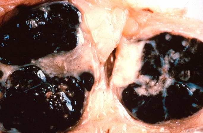
| 01:55, 21 August 2013 | IPLab7Melanoma2.jpg (file) |  |
36 KB | Seung Park | This is a gross photograph of lymph nodes almost entirely replaced by black pigment (melanin). | 1 |
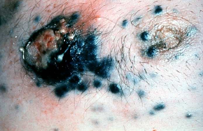
| 01:55, 21 August 2013 | IPLab7Melanoma1.jpg (file) |  |
52 KB | Seung Park | This is a gross photograph of skin with melanoma. Note the black pigment, multiple satellite nodules, and focal ulceration. Some of the satellite nodules affect the nipple. | 1 |
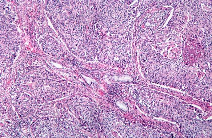
| 01:51, 21 August 2013 | IPLab7IDC8.jpg (file) |  |
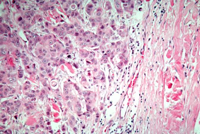
73 KB | Seung Park | This is a high-power photomicrograph demonstrating the growth pattern of the tumor. The tumor consists of malignant duct-lining cells growing in cords, solid cell nests, tubules, and glands. The cytologic detail of tumor cells varies from small cells w... | 1 |
| 01:51, 21 August 2013 | IPLab7IDC7.jpg (file) |  |
81 KB | Seung Park | This is a section taken at the periphery of the tumor showing bands of tumor cells infiltrating into the fat tissue. | 1 |
| 01:51, 21 August 2013 | IPLab7IDC6.jpg (file) |  |
70 KB | Seung Park | This is a section of breast tumor with abundant fibrous tissue throughout the tumor (desmoplasia, scirrhous carcinoma). | 1 |
| 01:51, 21 August 2013 | IPLab7IDC5.jpg (file) |  |
53 KB | Seung Park | This is a high-power photomicrograph showing the cellular and nuclear features of the tumor cells. The large epithelial cells form glands and are medium-sized with a moderate amount of cytoplasm, vesicular nuclei, and nucleoli. | 1 |
| 01:50, 21 August 2013 | IPLab7IDC4.jpg (file) |  |
63 KB | Seung Park | This is a higher-magnification showing abundant groups of tumor cells dissecting through the breast parenchyma - tumor infiltration (infiltrating duct cell carcinoma). | 1 |
| 01:50, 21 August 2013 | IPLab7IDC3.jpg (file) |  |
91 KB | Seung Park | This is a section of breast with small groups of carcinoma cells throughout the breast tissue and invading through the dermis. | 1 |
| 01:50, 21 August 2013 | IPLab7IDC2.jpg (file) |  |
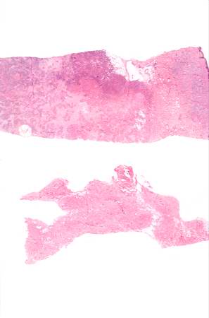
12 KB | Seung Park | These are sections of normal breast (lower) and breast tissue with infiltrating duct carcinoma (upper). Note the increased cellularity (increased blue staining due to the increased number of nuclei) in the tumor tissue. | 1 |
| 01:50, 21 August 2013 | IPLab7IDC1.jpg (file) |  |
56 KB | Seung Park | This is a gross photograph of the surgical specimen of breast with infiltrating duct carcinoma. Note the tumor tissue under the area of the nipple. The tumor infiltrates in an irregular fashion into the breast parenchyma. Note the nipple retraction cau... | 1 |
| 01:47, 21 August 2013 | IPLab7Metastatic9.jpg (file) |  |
78 KB | Seung Park | This is a high-power photomicrograph of the edge of the tumor nodule in the lung. The tumor cells area growing in a glandular pattern. The area of necrosis is evident at the right side of the image. | 1 |
| 01:47, 21 August 2013 | IPLab7Metastatic8.jpg (file) |  |
71 KB | Seung Park | This is a high-power photomicrograph of the edge of the tumor nodule in the lung. The tumor cells are infiltrating into the lung parenchyma (1). Even at this power you can see the glandular formation of this adenocarcinoma. There is a large area of nec... | 1 |
| 01:47, 21 August 2013 | IPLab7Metastatic7.jpg (file) |  |
71 KB | Seung Park | This is a photomicrograph of a tumor nodule in the lung. The tumor cells are infiltrating into the lung parenchyma (1). There is a large area of necrosis in the center of the tumor (2). | 1 |
| 01:46, 21 August 2013 | IPLab7Metastatic6.jpg (file) |  |
80 KB | Seung Park | This is a high-power photomicrograph of tumor cells that are forming glands (arrows). | 1 |
| 01:46, 21 August 2013 | IPLab7Metastatic5.jpg (file) |  |
85 KB | Seung Park | This is a higher-power photomicrograph showing how the tumor cells (arrows) have infiltrated into the liver parenchyma. | 1 |
| 01:46, 21 August 2013 | IPLab7Metastatic4.jpg (file) |  |
59 KB | Seung Park | This is a photomicrograph taken at the interface between the tumor (top) and the normal liver parenchyma (bottom). | 1 |
| 01:46, 21 August 2013 | IPLab7Metastatic3.jpg (file) |  |
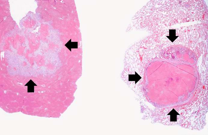
39 KB | Seung Park | These are low-power photomicrographs of a section of liver (left) and lung (right) containing tumor nodules (arrows). | 1 |
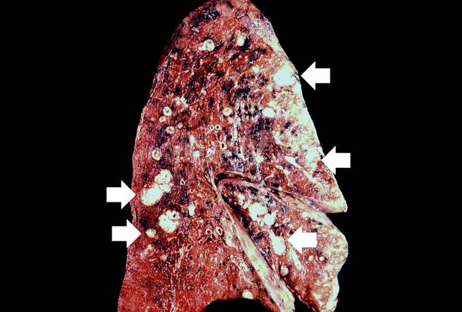
| 01:45, 21 August 2013 | IPLab7Metastatic2.jpg (file) |  |
45 KB | Seung Park | This gross photograph of the lung from this case also demonstrates multiple, variably sized pale/white-tan nodules scattered throughout the lung. | 1 |
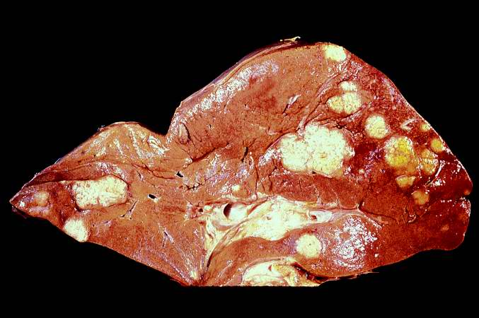
| 01:45, 21 August 2013 | IPLab7Metastatic1.jpg (file) |  |
50 KB | Seung Park | This gross photograph of the liver from this case demonstrates multiple, variably-sized pale/white-tan nodules scattered throughout the liver. | 1 |
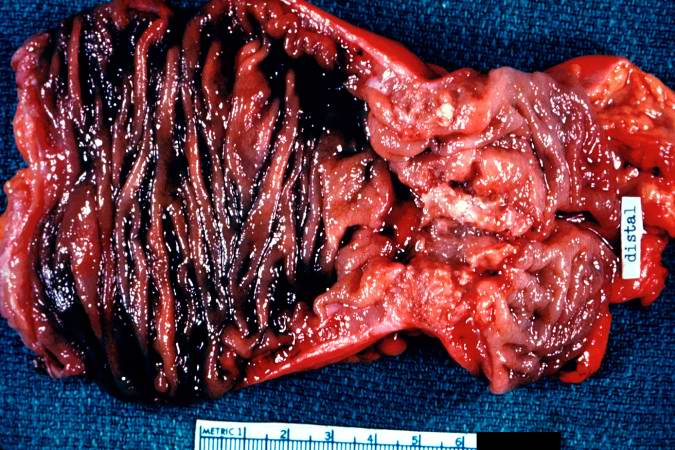
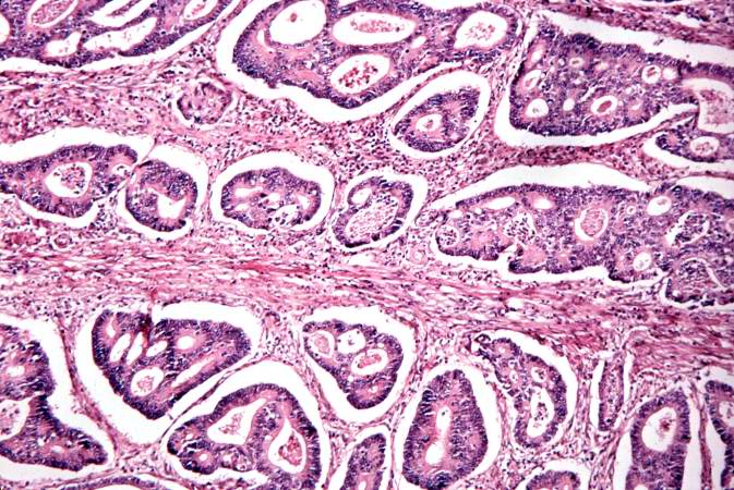
| 01:42, 21 August 2013 | IPLab7ColonCA9.jpg (file) |  |
83 KB | Seung Park | This is a segment of distal colon from another case. Note the annular tumor that severely compromises the lumen of the colon. There is dilation of the colon proximal to the tumor. | 1 |
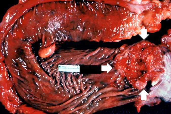
| 01:42, 21 August 2013 | IPLab7ColonCA8.jpg (file) |  |
66 KB | Seung Park | This gross photograph from another case demonstrates an ulcerated adenocarcinoma (arrows) at the rectosigmoid junction. | 1 |
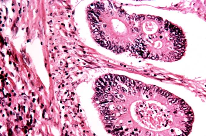
| 01:42, 21 August 2013 | IPLab7ColonCA7.jpg (file) |  |
65 KB | Seung Park | This is a high-power photomicrograph of tumor cells forming glands. | 1 |
| 01:41, 21 August 2013 | IPLab7ColonCA6.jpg (file) |  |
96 KB | Seung Park | This is a high-power photomicrograph of tumor cells forming glands. | 1 |