File list
This special page shows all uploaded files.
| Date | Name | Thumbnail | Size | Description | Versions |
|---|---|---|---|---|---|
| 01:40, 21 August 2013 | IPLab7ColonCA1.jpg (file) |  |
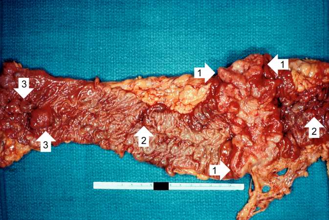
69 KB | This is a gross photograph of the adenoma from the surgical specimen in this case. Note the large, ulcerated, fungating annular (encircling) carcinoma (1) with areas of hemorrhage (2). Also note the adenomatous polyps (3). | 1 |
| 01:37, 21 August 2013 | IPLab7EsophSCC7.jpg (file) |  |
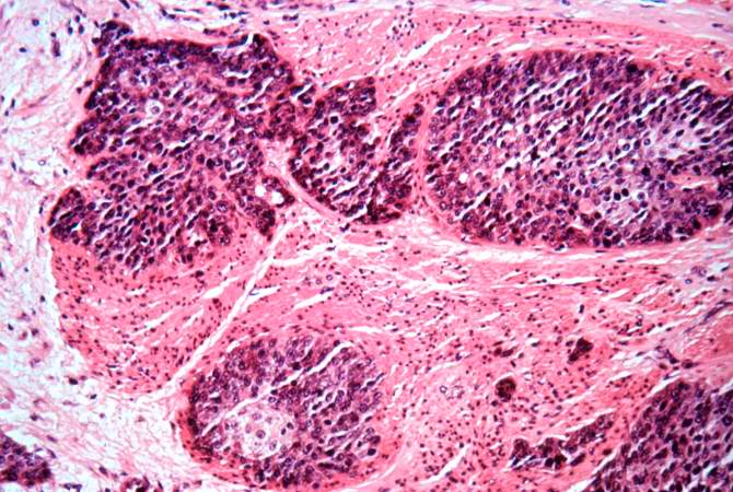
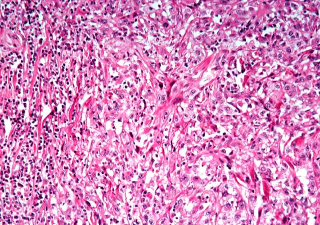
81 KB | This is a high-power photomicrograph of the tumor cells that have invaded the adjacent muscle tissue. | 1 |
| 01:37, 21 August 2013 | IPLab7EsophSCC6.jpg (file) |  |
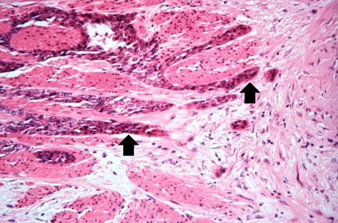
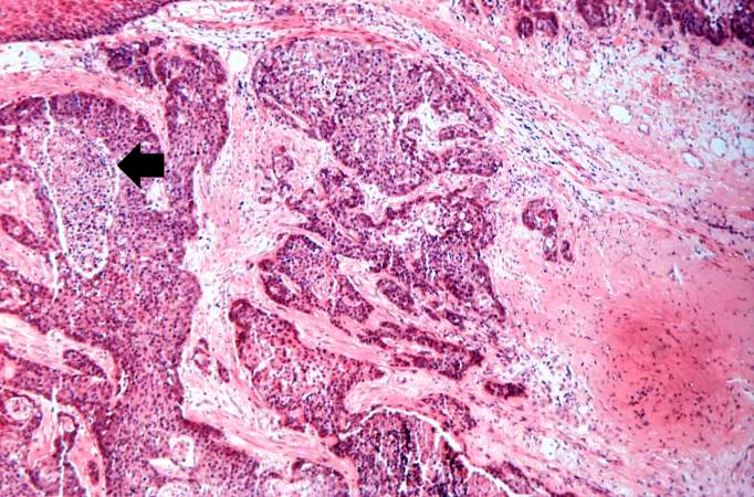
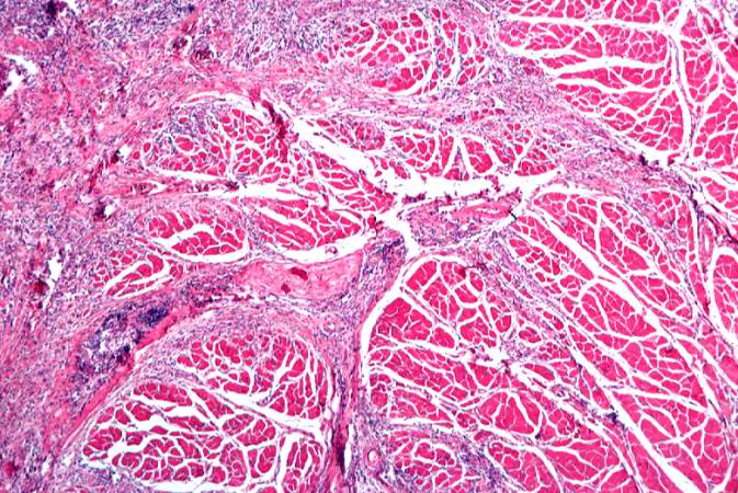
67 KB | This is a higher-power photomicrograph of bands of tumor cells (arrows) extending between the muscle bundles. | 1 |
| 01:37, 21 August 2013 | IPLab7EsophSCC5.jpg (file) |  |
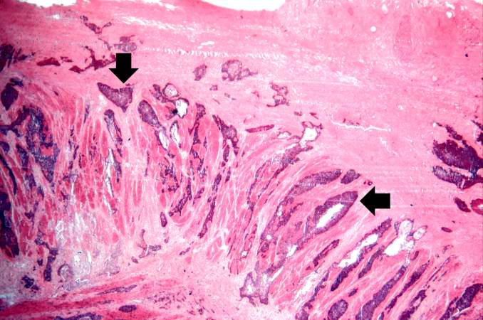
63 KB | This is a photomicrograph of bands of tumor cells invading into the adjacent tissues (arrows). | 1 |
| 01:37, 21 August 2013 | IPLab7EsophSCC4.jpg (file) |  |
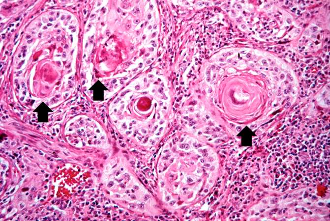
86 KB | This is a higher-power photomicrograph showing invasive squamous cell carcinoma. Tongues and islands of tumor cells exhibit areas of central necrosis (arrow). | 1 |
| 01:36, 21 August 2013 | IPLab7EsophSCC3.jpg (file) |  |
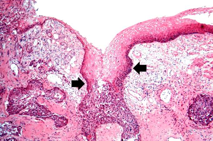
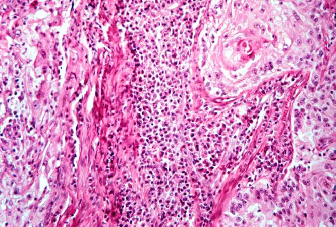
74 KB | This is a high-power photomicrograph demonstrating the normal epithelium undergoing transition to carcinoma (arrows). | 1 |
| 01:36, 21 August 2013 | IPLab7EsophSCC2.jpg (file) |  |
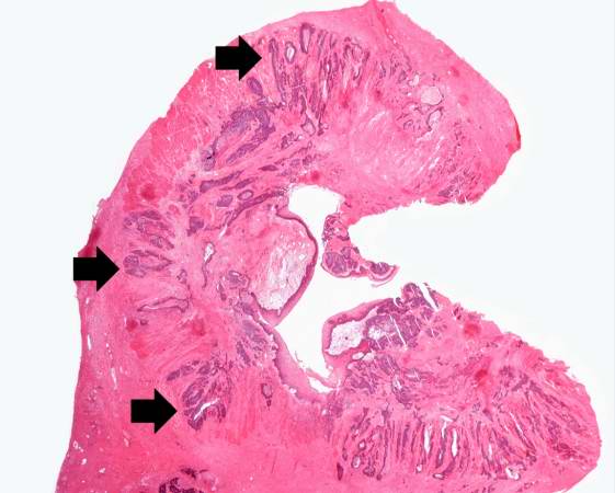
34 KB | This low-power photomicrograph of a cross-section through the esophagus at the area of constriction shows extensive infiltration of the esophageal wall with squamous cell carcinoma (arrows). | 1 |
| 01:36, 21 August 2013 | IPLab7EsophSCC1.jpg (file) |  |
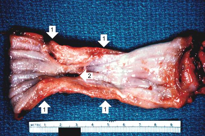
66 KB | This is a gross photograph of the luminal surface of the esophagus with the area of constriction (1). The area protrudes into the lumen. There is also a central area of ulceration (2). | 1 |
| 01:33, 21 August 2013 | IPLab7LipSCC8.jpg (file) |  |
101 KB | This is a section of muscle tissue from this biopsy of the lip. Note that the squamous cell carcinoma has infiltrated into the muscle tissue. There are also inflammatory cells within this area of tumor infiltration. | 1 |
| 01:33, 21 August 2013 | IPLab7LipSCC7.jpg (file) |  |
85 KB | This is a high power photomicrograph of a poorly-differentiated area of tumor. Note the spindle-shaped cells and the irregular pattern of growth. | 1 |
| 01:32, 21 August 2013 | IPLab7LipSCC6.jpg (file) |  |
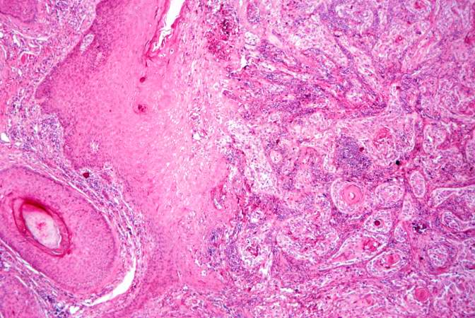
85 KB | This is a high power photomicrograph of the well-differentiated squamous cell carcinoma. Note the intracytoplasmic keratinization which gives the cells a glassy appearance. The focal accumulations of keratinized cells are called keratin pearls (arrows). | 1 |
| 01:32, 21 August 2013 | IPLab7LipSCC5.jpg (file) |  |
79 KB | This is a higher-power photomicrograph of infiltrating squamous cell carcinoma and inflammatory cells. | 1 |
| 01:32, 21 August 2013 | IPLab7LipSCC4.jpg (file) |  |
77 KB | This is a higher-power photomicrograph of the well-differentiated squamous cell carcinoma and the inflammatory cell infiltration. | 1 |
| 01:32, 21 August 2013 | IPLab7LipSCC3.jpg (file) |  |
57 KB | This photomicrograph shows a large area of ulceration (arrow) with underlying congestion and hemorrhage. The area of ulceration is adjacent to an area of tumor infiltration. | 1 |
| 01:31, 21 August 2013 | IPLab7LipSCC2.jpg (file) |  |
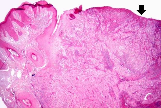
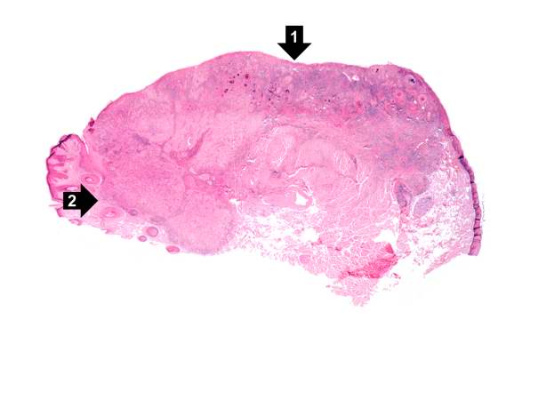
21 KB | This is a low-power photomicrograph of squamous cell carcinoma of the lip. Note focal ulceration (1) and tumor infiltration at the vermilion border (2). | 1 |
| 01:31, 21 August 2013 | IPLab7LipSCC1.jpg (file) |  |
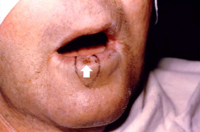
30 KB | This is a pre-op photograph of this patient with an ulcerated lesion on his lip (arrow). Also note that the lip is somewhat thickened. The area for surgical excision is delineated by black marker. | 1 |
| 01:28, 21 August 2013 | IPLab7Fibroadenoma7.jpg (file) |  |
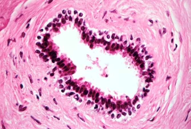
49 KB | This is a higher magnification of fibroadenoma showing irregularly shaped ducts lined by two layers of cells as previously described. | 1 |
| 01:28, 21 August 2013 | IPLab7Fibroadenoma6.jpg (file) |  |
61 KB | This is a high magnification of the fibroadenoma showing the dense stroma of the tumor surrounding the irregularly shaped duct. The ducts are lined by two cell layers, one of cuboidal, two columnar cells (inner layer) and an outer layer of flattened ce... | 1 |
| 01:28, 21 August 2013 | IPLab7Fibroadenoma5.jpg (file) |  |
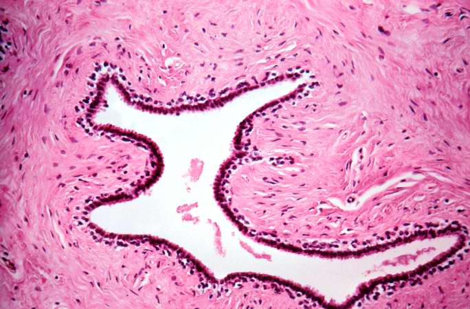
68 KB | This is a higher-power photomicrograph of fibroadenoma showing ducts embedded in connective tissue. | 1 |
| 01:27, 21 August 2013 | IPLab7Fibroadenoma4.jpg (file) |  |
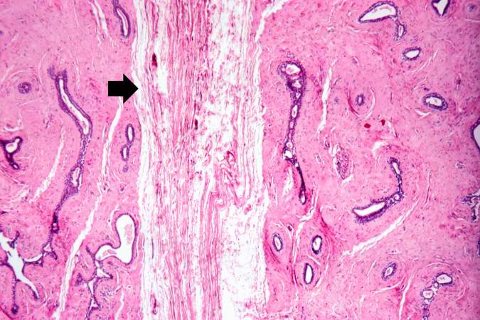
67 KB | This photomicrograph shows the compressed connective tissue (arrow) between two nodules of dense fibrous tissue and ducts. | 1 |
| 01:27, 21 August 2013 | IPLab7Fibroadenoma3.jpg (file) |  |
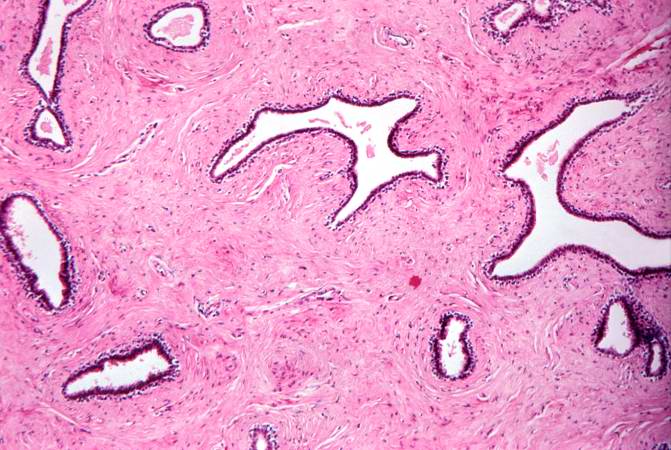
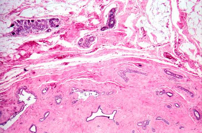
67 KB | This is a higher magnification of the fibroadenoma showing the dense stroma of the tumor surrounding the irregularly shaped ducts. The adjacent fibrofatty tissue containing breast ducts and lobules has been compressed by the tumor. | 1 |
| 01:27, 21 August 2013 | IPLab7Fibroadenoma2.jpg (file) |  |
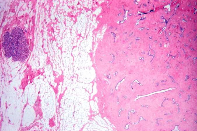
55 KB | This is a higher magnification of one of the three nodules. At this power, the nodule seems to be composed of a solid parenchyma with small glandular spaces. The adjacent breast parenchyma consists mostly of fat. | 1 |
| 01:26, 21 August 2013 | IPLab7Fibroadenoma1.jpg (file) |  |
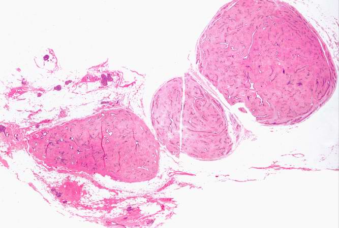
43 KB | This low-power photomicrograph of the surgical specimen demonstrates three ovoid, well-circumscribed nodules surrounded by fibroadipose tissue. | 1 |
| 01:23, 21 August 2013 | IPLab7Adenoma6.jpg (file) |  |
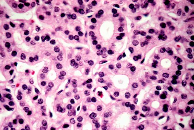
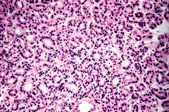
57 KB | This high-power photomicrograph demonstrates the relatively normal cellular morphology of this follicular adenoma. | 1 |
| 01:23, 21 August 2013 | IPLab7Adenoma5.jpg (file) |  |
88 KB | This is a photomicrograph of an adenoma. Note that the follicular architecture is well developed and more or less uniform throughout this section. | 1 |
| 01:23, 21 August 2013 | IPLab7Adenoma4.jpg (file) |  |
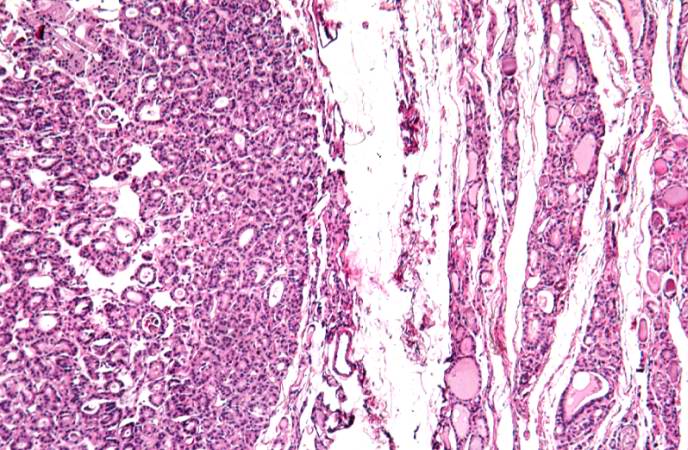
61 KB | This photomicrograph demonstrates the densely packed follicular pattern in the adenoma (left) and the larger colloid-filled follicles of the normal thyroid (right). An area of compressed thyroid is present adjacent to the adenoma (arrows). | 1 |
| 01:22, 21 August 2013 | IPLab7Adenoma3.jpg (file) |  |
89 KB | This is another higher-power photomicrograph of the adenoma (left) and the adjacent thyroid tissue (right). Note the compression of the adjacent normal thyroid and the difference in morphology between the adenoma and the thyroid. | 1 |
| 01:22, 21 August 2013 | IPLab7Adenoma2.jpg (file) |  |
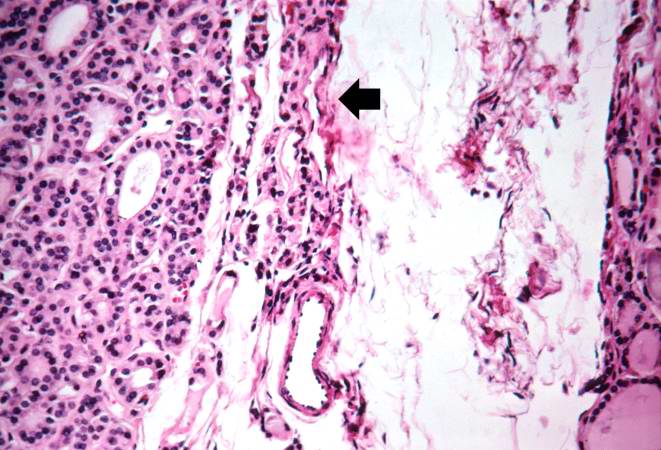
72 KB | This is a higher-power view of the border between the tumor mass and the adjacent thyroid tissue. Note that the mass has compressed the adjacent normal thyroid tissue (arrow). Also note the different morphology between the adenoma (very cellular, dense... | 1 |
| 01:22, 21 August 2013 | IPLab7Adenoma1.jpg (file) |  |
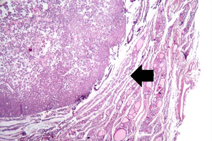
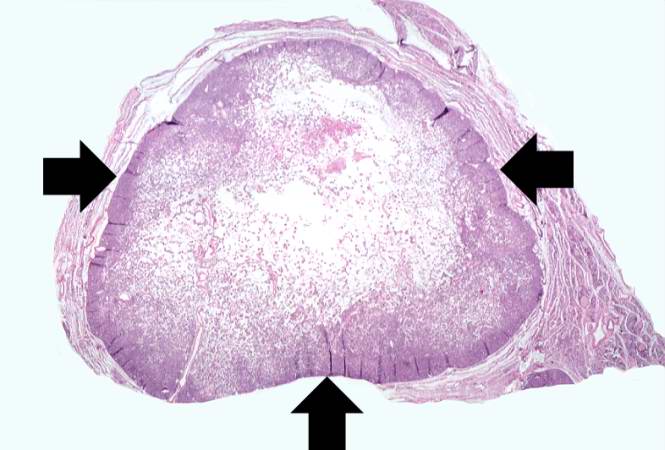
42 KB | This is a low-power photomicrograph of a nodule found in the thyroid of this case. Note that the mass is well-circumscribed and there is a sharp line of demarcation between the mass and the adjacent thyroid tissue (arrows). | 1 |
| 21:58, 20 August 2013 | IPLab6AcuteRejection10.jpg (file) |  |
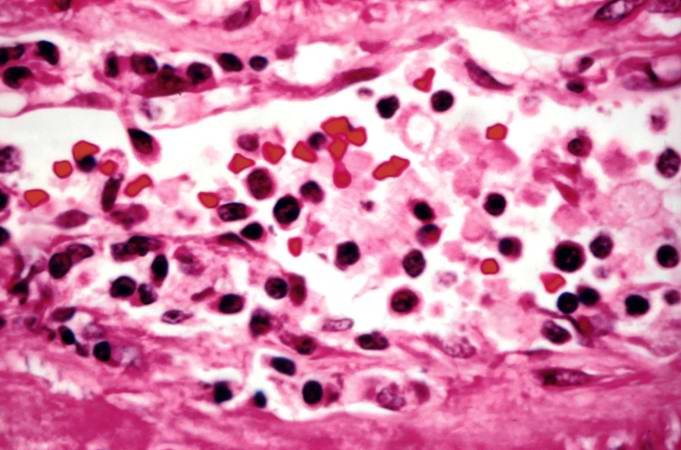
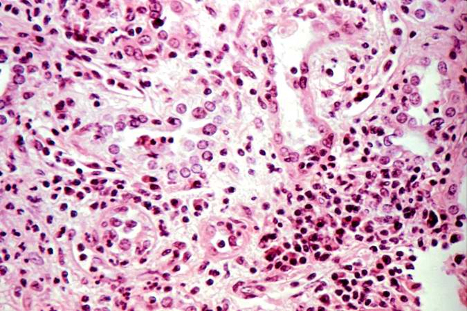
75 KB | This high-power photomicrograph demonstrates the cellular infiltrate within the interstitium and cells within the renal tubules. | 1 |
| 21:58, 20 August 2013 | IPLab6AcuteRejection9.jpg (file) |  |
47 KB | This is a high-power photomicrograph of cells infiltrating the wall of the blood vessel. | 1 |
| 21:58, 20 August 2013 | IPLab6AcuteRejection8.jpg (file) |  |
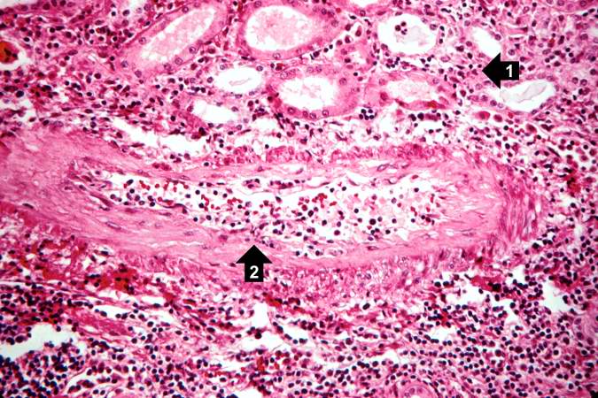
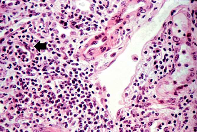
88 KB | This high-power photomicrograph demonstrates the cellular infiltrate within the interstitium (1) and in the wall of the blood vessel (2). | 1 |
| 21:57, 20 August 2013 | IPLab6AcuteRejection7.jpg (file) |  |
74 KB | This high-power photomicrograph demonstrates the cellular infiltrate within the interstitium and in the wall of the blood vessel on the left. | 1 |
| 21:57, 20 August 2013 | IPLab6AcuteRejection6.jpg (file) |  |
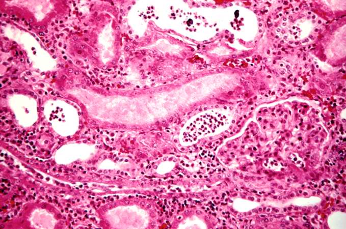
95 KB | This is a higher-power photomicrograph demonstrating the cellular infiltrate within the interstitium. There is some degeneration (coagulative necrosis) of tubules and glomeruli. | 1 |
| 21:57, 20 August 2013 | IPLab6AcuteRejection5.jpg (file) |  |
83 KB | This is a higher-power photomicrograph demonstrating the cellular infiltrate within the interstitium and around the small blood vessel in the center of the image. | 1 |
| 21:57, 20 August 2013 | IPLab6AcuteRejection4.jpg (file) |  |
78 KB | This is a higher-power photomicrograph demonstrating the cellular infiltrates within this kidney section. Note that in addition to the diffuse cellularity, the focal accumulations of cells appear to be focused around blood vessels. | 1 |
| 21:56, 20 August 2013 | IPLab6AcuteRejection3.jpg (file) |  |
69 KB | This is a higher-power photomicrograph demonstrating the cellular infiltrates within this kidney section. | 1 |
| 21:56, 20 August 2013 | IPLab6AcuteRejection2.jpg (file) |  |
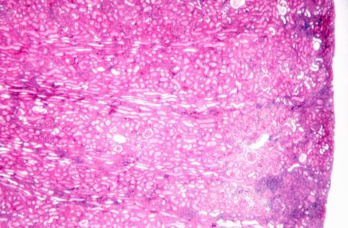
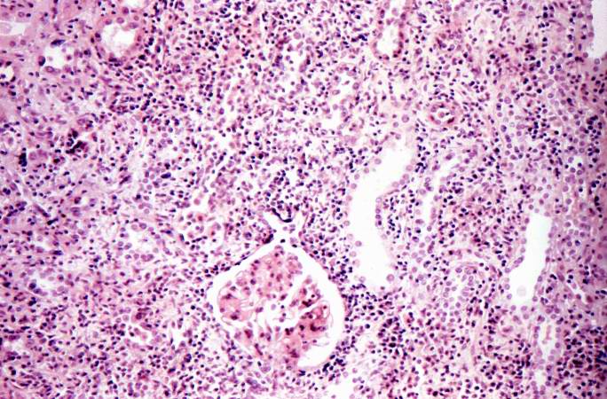
41 KB | This is a low-power photomicrograph of the kidney that was removed from this patient. Even at this low power you can appreciate the focal accumulations of cells within this section and the diffuse cellular infiltrate (blue dots) throughout the kidney p... | 1 |
| 21:56, 20 August 2013 | IPLab6AcuteRejection1.jpg (file) |  |
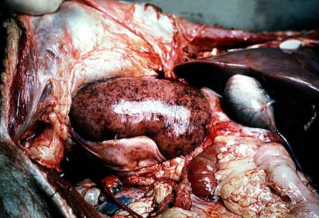
66 KB | This is a gross photograph of a kidney with acute rejection from an autopsy case. Note that the kidney is swollen (edema and inflammation) and there are areas of hemorrhage throughout the kidney. | 1 |
| 21:51, 20 August 2013 | IPLab6ChronicRejection12.jpg (file) |  |
72 KB | Photomicrograph from another region of previous image. Note the cellular infiltrate around a small blood vessel (right) and neutrophils within renal tubules (arrow). | 1 |
| 21:51, 20 August 2013 | IPLab6ChronicRejection11.jpg (file) |  |
71 KB | This is a higher-power photomicrograph of kidney from the previous image demonstrating the cellular infiltrate which is comprised of lymphocytes, macrophages, plasma cells and a few neutrophils. | 1 |
| 21:50, 20 August 2013 | IPLab6ChronicRejection10.jpg (file) |  |
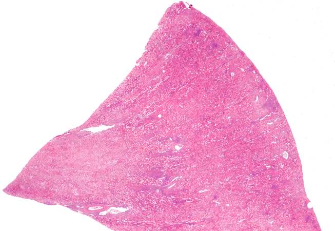
86 KB | This is a high-power photomicrograph of a kidney from another case of chronic transplant rejection. In this case there is extensive damage to the kidney due to the chronic rejection (loss of tubules and glomerular lesions). In addition, this kidney was... | 1 |
| 21:50, 20 August 2013 | IPLab6ChronicRejection9.jpg (file) |  |
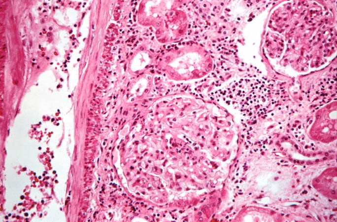
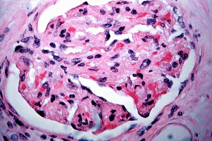
61 KB | This is a high-power photomicrograph of a damaged glomerulus. Note the loss of normal capillary structure, the mesangial expansion and the infiltration of large mononuclear cells. | 1 |
| 21:50, 20 August 2013 | IPLab6ChronicRejection8.jpg (file) |  |
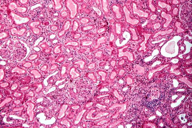
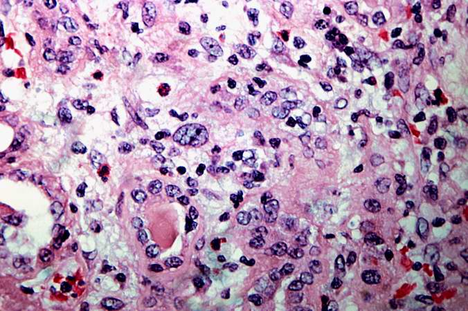
73 KB | This is a high-power photomicrograph of renal cortex with cellular infiltrate and few remaining renal tubules. The cellular infiltrate comprises macrophages, activated (large) lymphocytes and a few neutrophils and plasma cells. | 1 |
| 21:49, 20 August 2013 | IPLab6ChronicRejection7.jpg (file) |  |
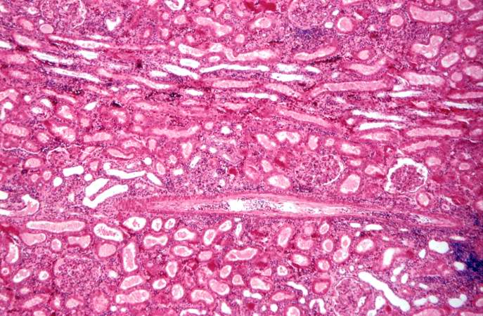
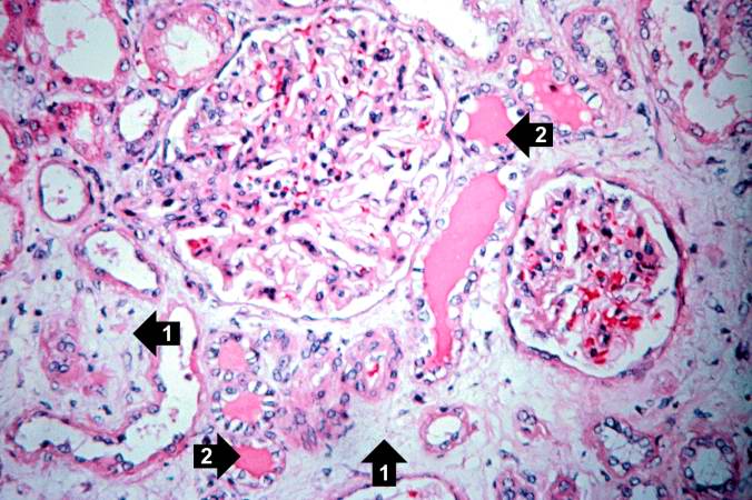
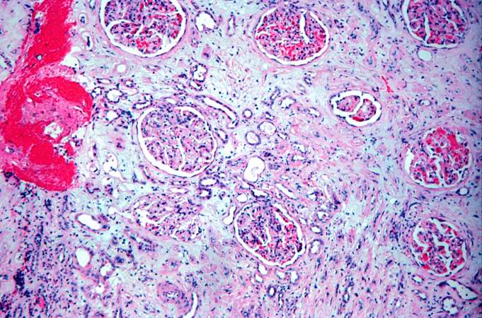
70 KB | This is a photomicrograph of a glomerulus with a mild cellular infiltrate (left) and a small damaged glomerulus (right). There is extensive interstitial fibrosis (1), loss of renal tubules, and the remaining tubules contain protein (2) indicating sever... | 1 |
| 21:49, 20 August 2013 | IPLab6ChronicRejection6.jpg (file) |  |
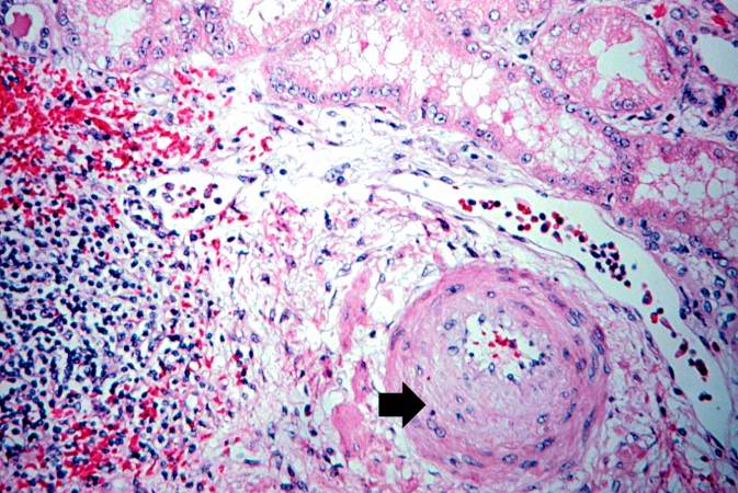
81 KB | This is a photomicrograph of rejected kidney with a focus of cellular infiltrate (left) and a small artery with neointimal proliferation and stenosis (arrow). | 1 |
| 21:49, 20 August 2013 | IPLab6ChronicRejection5.jpg (file) |  |
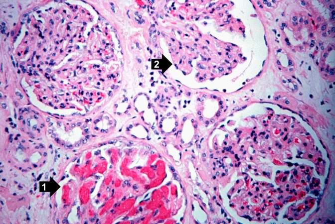
71 KB | This high-power photomicrograph of glomeruli from this kidney demonstrates congestion (1), increased cellularity of glomeruli with mesangial expansion, and a glomerulus that is almost completely obliterated or sclerosed (2). | 1 |
| 21:49, 20 August 2013 | IPLab6ChronicRejection4.jpg (file) |  |
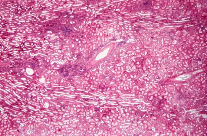
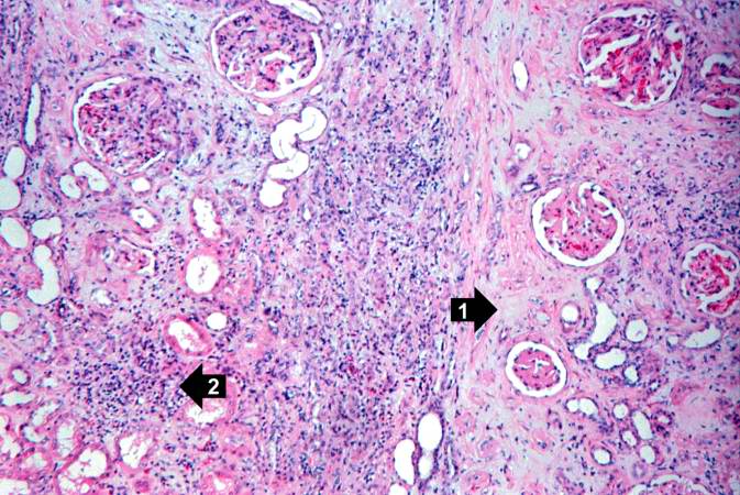
84 KB | This is another area of renal cortex similar to the previous image. Note the fibrosis (1) and loss of renal tubules throughout this section. Also note the focus of inflammatory cells (2) indicating that despite the chromic nature of this lesion, there ... | 1 |
| 21:48, 20 August 2013 | IPLab6ChronicRejection3.jpg (file) |  |
89 KB | This is a photomicrograph of kidney with a focal area of hemorrhage around a small blood vessel (left) and congestion of the glomeruli. Note that there is a marked loss of renal tubules throughout this section with replacement by fibrous connective tis... | 1 |
| 21:48, 20 August 2013 | IPLab6ChronicRejection2.jpg (file) |  |
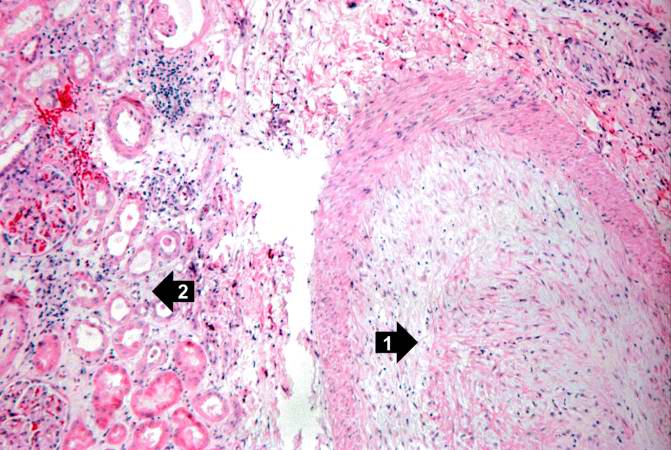
68 KB | This is a higher-power photomicrograph of kidney containing a section of blood vessel that demonstrates a marked neointimal proliferative response (1). In this case the lumen of the artery is obliterated. Also note the cellular infiltrate in the inters... | 1 |