File list
This special page shows all uploaded files.
| Date | Name | Thumbnail | Size | User | Description | Versions |
|---|---|---|---|---|---|---|
| 21:49, 20 August 2013 | IPLab6ChronicRejection5.jpg (file) |  |
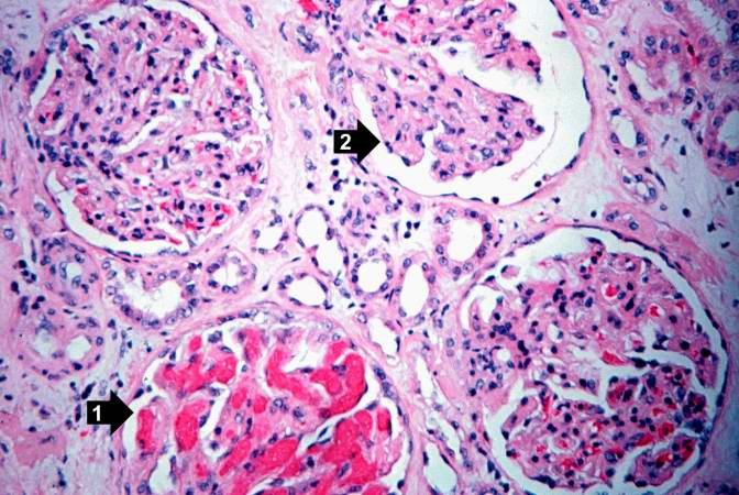
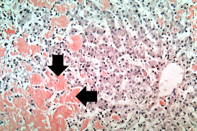
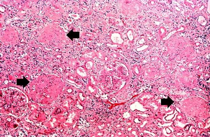
71 KB | Seung Park | This high-power photomicrograph of glomeruli from this kidney demonstrates congestion (1), increased cellularity of glomeruli with mesangial expansion, and a glomerulus that is almost completely obliterated or sclerosed (2). | 1 |
| 21:49, 20 August 2013 | IPLab6ChronicRejection4.jpg (file) |  |
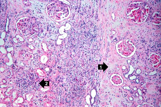
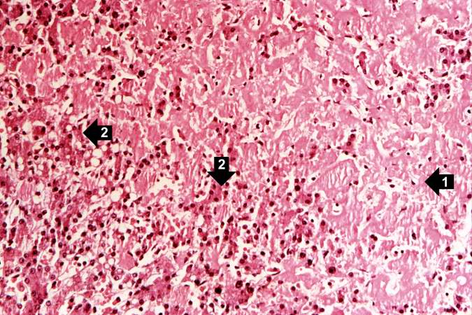
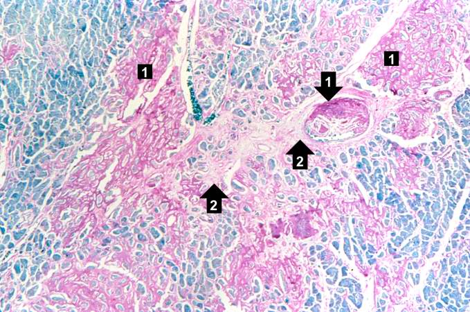
84 KB | Seung Park | This is another area of renal cortex similar to the previous image. Note the fibrosis (1) and loss of renal tubules throughout this section. Also note the focus of inflammatory cells (2) indicating that despite the chromic nature of this lesion, there ... | 1 |
| 21:48, 20 August 2013 | IPLab6ChronicRejection3.jpg (file) |  |
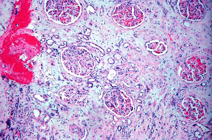
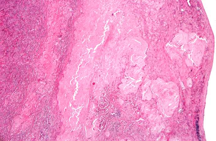
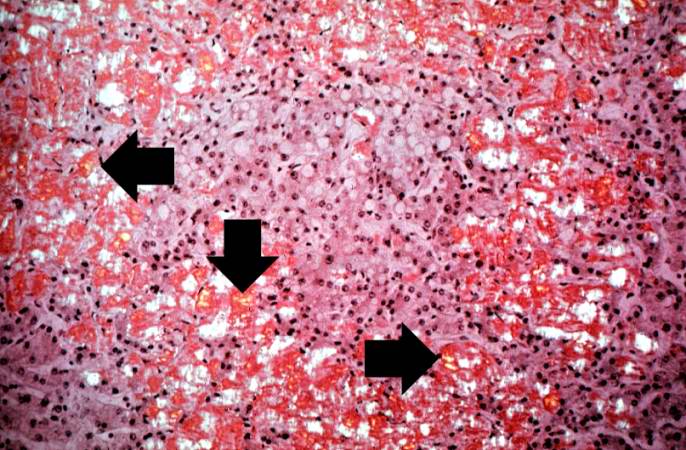
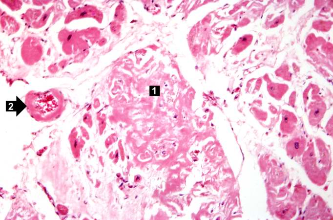
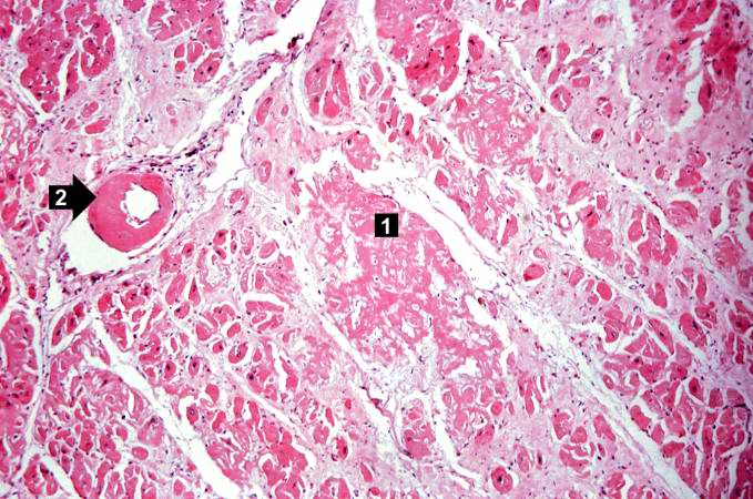
89 KB | Seung Park | This is a photomicrograph of kidney with a focal area of hemorrhage around a small blood vessel (left) and congestion of the glomeruli. Note that there is a marked loss of renal tubules throughout this section with replacement by fibrous connective tis... | 1 |
| 21:48, 20 August 2013 | IPLab6ChronicRejection2.jpg (file) |  |
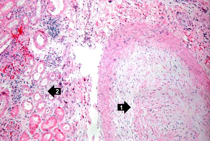
68 KB | Seung Park | This is a higher-power photomicrograph of kidney containing a section of blood vessel that demonstrates a marked neointimal proliferative response (1). In this case the lumen of the artery is obliterated. Also note the cellular infiltrate in the inters... | 1 |
| 21:48, 20 August 2013 | IPLab6ChronicRejection1.jpg (file) |  |
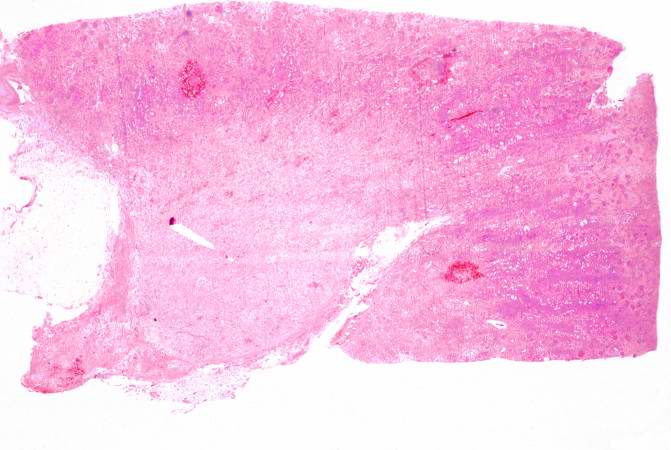
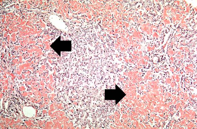
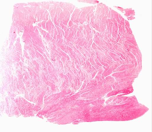
43 KB | Seung Park | This is a low-power photomicrograph of the kidney from this case of chronic transplant rejection. Note the focal areas of hemorrhage and inflammatory cell infiltrate in this section. | 1 |
| 21:43, 20 August 2013 | IPLab6MM4.jpg (file) |  |
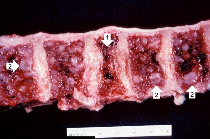
47 KB | Seung Park | This is a photograph of the vertebral column from this patient at autopsy. Notice the collapsed vertebra (1). There are multiple variably-sized white nodules (2) within the bone marrow. These are accumulations of malignant plasma cells in this case of ... | 1 |
| 21:43, 20 August 2013 | IPLab6MM3.jpg (file) |  |
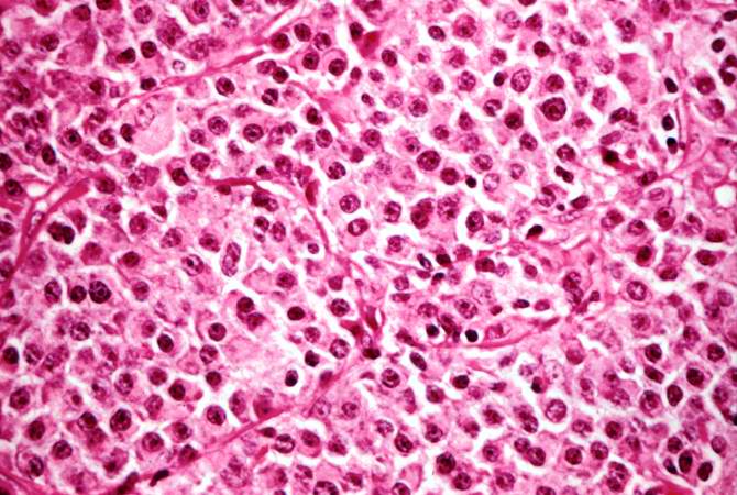
72 KB | Seung Park | This high-power photomicrograph demonstrates the cells that make up this tissue. These cells resemble plasma cells and are the malignant cell of multiple myeloma. | 1 |
| 21:42, 20 August 2013 | IPLab6MM2.jpg (file) |  |
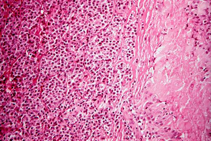
81 KB | Seung Park | This higher-power photomicrograph shows the junction between an amorphous hyaline-appearing area (amyloid) on the right and cellular areas (plasmacytoid cells) on the left. | 1 |
| 21:42, 20 August 2013 | IPLab6MM1.jpg (file) |  |
62 KB | Seung Park | This is a low-power photomicrograph of the mediastinal mass. The mass is encapsulated and contains cellular areas (blue) and areas of pale red material. | 1 |
| 21:36, 20 August 2013 | IPLab6Amyloid11.jpg (file) |  |
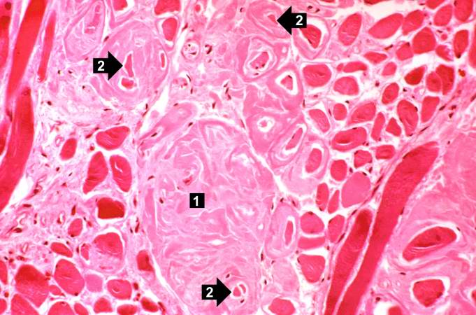
58 KB | Seung Park | This photomicrograph of the tongue demonstrates extensive amyloid deposits (1) separating the skeletal muscle fibers of the tongue. In many cases the amyloid encircles the muscle fibers (2) and these muscle fibers are atrophied. | 1 |
| 21:36, 20 August 2013 | IPLab6Amyloid10.jpg (file) |  |
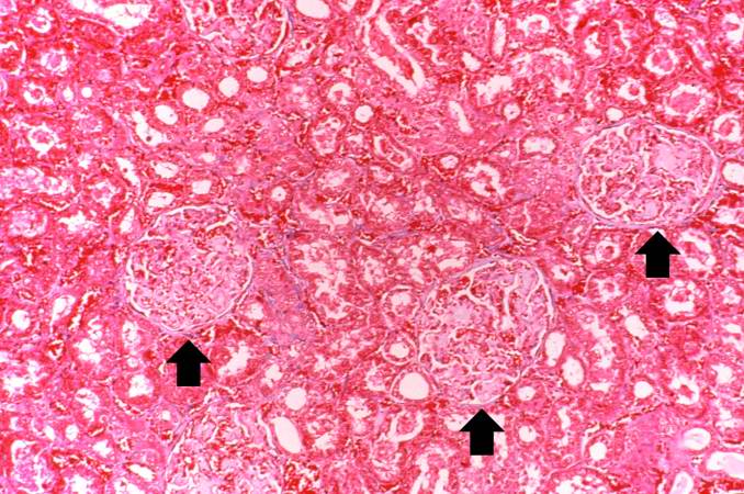
85 KB | Seung Park | This photomicrograph of kidney demonstrates the amyloid deposits (arrows) within glomeruli. | 1 |
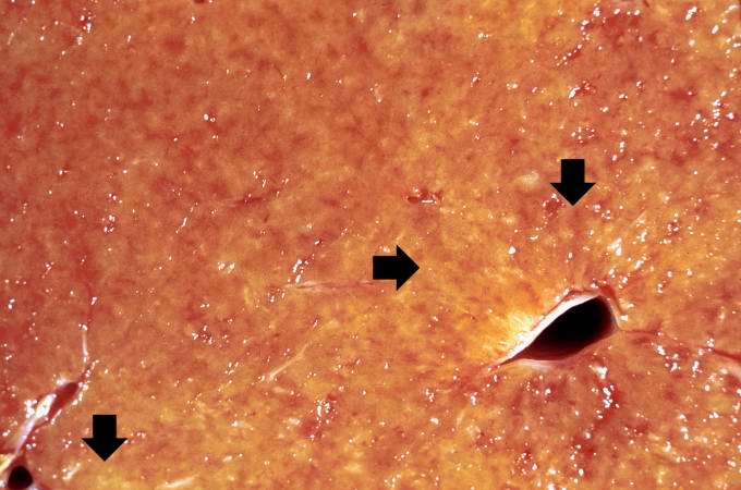
| 21:36, 20 August 2013 | IPLab6Amyloid9.jpg (file) |  |
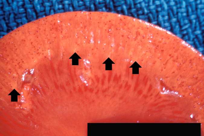
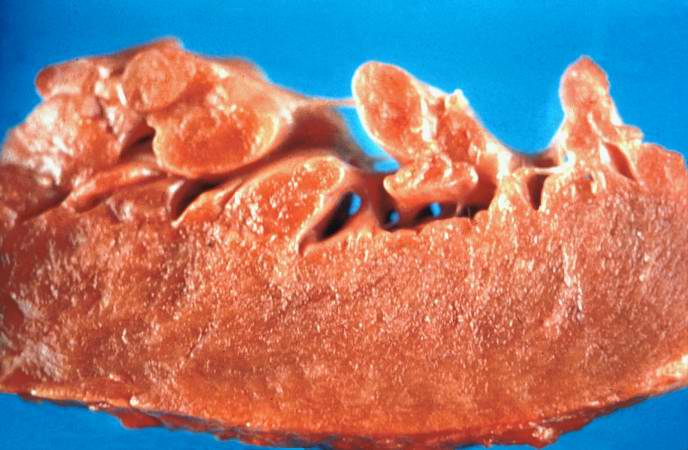
34 KB | Seung Park | This is a gross photograph of kidney from this case. Note the pale yellow material within the cortex (arrows). This is indicative of amyloid within the cortex and the glomeruli. Also note that there are multiple red spots in the cortex. These represent... | 1 |
| 21:35, 20 August 2013 | IPLab6Amyloid8.jpg (file) |  |
78 KB | Seung Park | This is a photomicrograph of Congo-red-stained liver tissue viewed with partially polarized light. Although not well demonstrated in this image, Congo-red-stained amyloid viewed through polarized light should give off a classic “apple green” birefr... | 1 |
| 21:35, 20 August 2013 | IPLab6Amyloid7.jpg (file) |  |
75 KB | Seung Park | This is a high-power view of liver tissue stained with Congo red. The orange amyloid material (arrows) is seen clearly between liver parenchymal cells. | 1 |
| 21:35, 20 August 2013 | IPLab6Amyloid6.jpg (file) |  |
92 KB | Seung Park | This is a low-power photomicrograph of liver tissue stained with Congo red (orange color in slide). Congo red reacts with amyloid and gives it an orange color (arrows). | 1 |
| 21:34, 20 August 2013 | IPLab6Amyloid5.jpg (file) |  |
85 KB | Seung Park | This is a higher-power photomicrograph showing the amyloid deposits (1) between hepatocytes (2). | 1 |
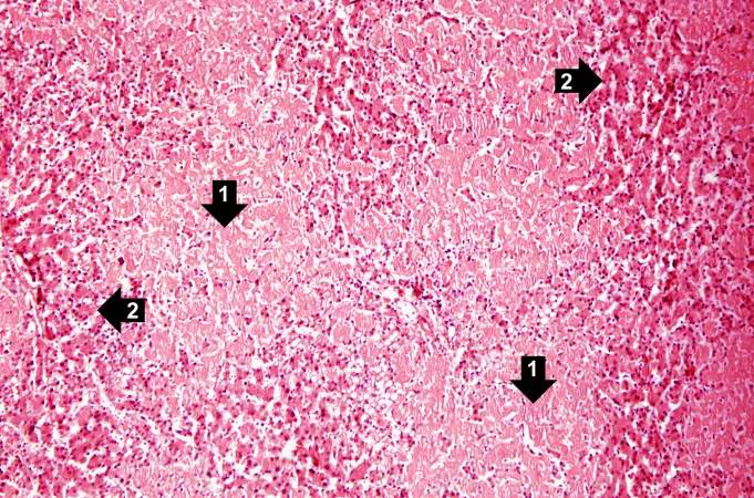
| 21:34, 20 August 2013 | IPLab6Amyloid4.jpg (file) |  |
97 KB | Seung Park | This is a low-power photomicrograph of liver tissue from this case. Note the eosinophilic hyaline material (1) present within and between the hepatic tissue (2). There is marked distortion of lobular architecture by the amyloid. | 1 |
| 21:34, 20 August 2013 | IPLab6Amyloid3.jpg (file) |  |
47 KB | Seung Park | This is a closer view of the cut surface of this liver. The pale waxy material can be seen within the hepatic tissue (arrows). | 1 |
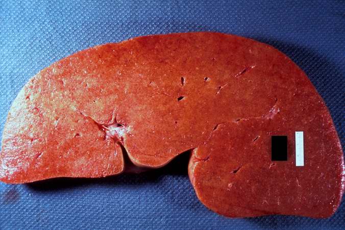
| 21:33, 20 August 2013 | IPLab6Amyloid2.jpg (file) |  |
60 KB | Seung Park | This is a gross picture of the cut surface of the liver from this case. The liver tissue is firm and has a waxy appearance--although this is difficult to appreciate in an image. | 1 |
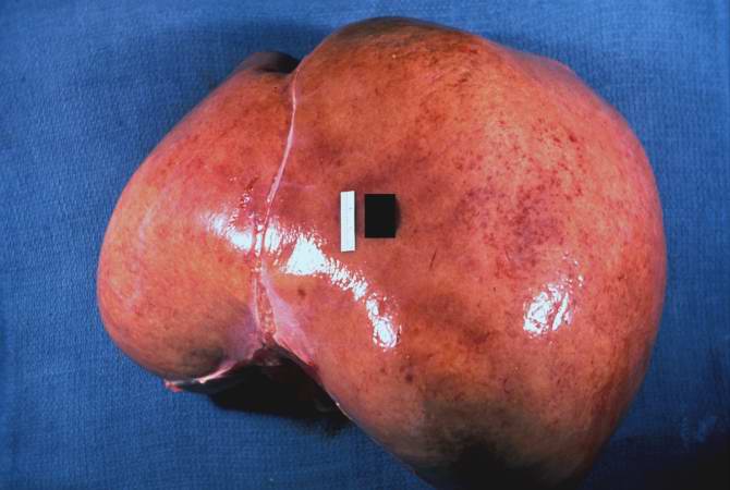
| 21:33, 20 August 2013 | IPLab6Amyloid1.jpg (file) |  |
35 KB | Seung Park | This is a gross picture of liver from this case. Note the pale, swollen appearance of this liver. | 1 |
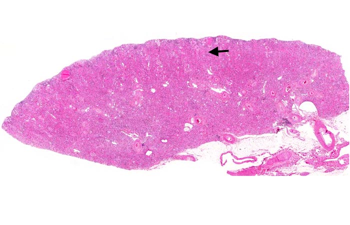
| 20:43, 20 August 2013 | IPLab6GN1.jpg (file) |  |
72 KB | Peter Anderson | This is a low-power photomicrograph of a saggital section of end stage chronic glomerulonephritis (GN). Note the marked thinning of the cortex (arrow). | 1 |
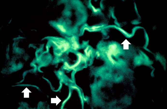
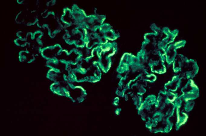
| 20:30, 20 August 2013 | IPLab6GN10.jpg (file) |  |
29 KB | Peter Anderson | For comparison this is an immunofluorescent photomicrograph of a glomerulus from a patient with Goodpasture's syndrome. The linear (arrows) immunofluorescence is characteristic of Goodpasture's syndrome. | 1 |
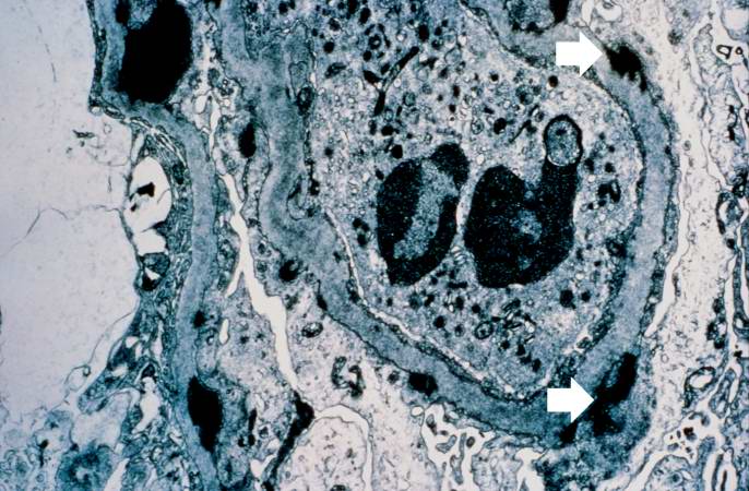
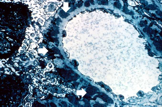
| 20:30, 20 August 2013 | IPLab6GN9.jpg (file) |  |
74 KB | Peter Anderson | This electron micrograph demonstrates scattered subepithelial dense deposits (arrows) and a polymorphonuclear leukocyte in the lumen. | 1 |
| 20:30, 20 August 2013 | IPLab6GN8.jpg (file) |  |
31 KB | Peter Anderson | This immunofluorescent photomicrograph of a glomerulus from a case of acute poststreptococcal glomerulonephritis shows a granular immunofluorescence pattern consistent with immune complex disease. The primary antibody used for this staining was specifi... | 1 |
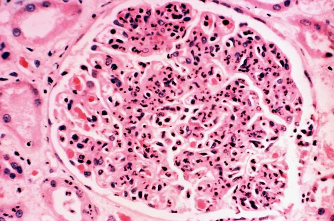
| 20:29, 20 August 2013 | IPLab6GN7.jpg (file) |  |
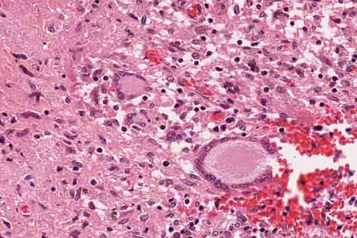
62 KB | Peter Anderson | This is a photomicrograph of a glomerulus from another case with acute poststreptococcal glomerulonephritis. In this case the immune complex glomerular disease is ongoing with necrosis and accumulation of neutrophils in the glomerulus. | 1 |
| 20:29, 20 August 2013 | IPLab6GN6.jpg (file) |  |
66 KB | Peter Anderson | This is an electron micrograph of subepithelial granular electron dense deposits (arrows) which correspond to the granular immunofluorescence seen in the previous image. | 1 |
| 20:28, 20 August 2013 | IPLab6GN5.jpg (file) |  |
30 KB | Peter Anderson | This is an immunofluorescent photomicrograph of granular membranous immunofluorescence (immune complex disease). The antibody used for these studies was specific for IgG. | 1 |
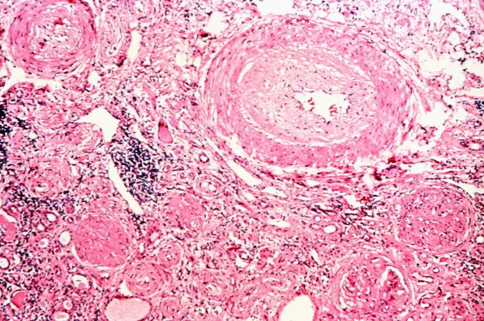
| 20:28, 20 August 2013 | IPLab6GN4.jpg (file) |  |
87 KB | Peter Anderson | This is a photomicrograph of interstitial and vascular lesions in end stage renal disease. | 1 |
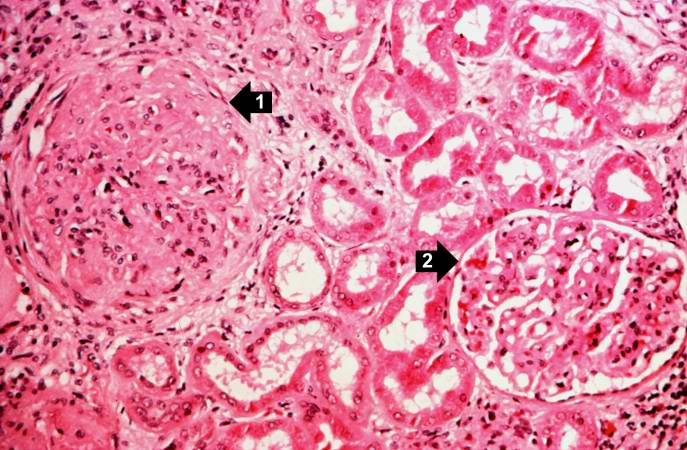
| 20:27, 20 August 2013 | IPLab6GN3.jpg (file) |  |
71 KB | Peter Anderson | This is a higher-power photomicrograph of hyalinized glomeruli (1) and glomeruli with thickened basement membranes (2). | 1 |
| 20:27, 20 August 2013 | IPLab6GN2.jpg (file) |  |
103 KB | Peter Anderson | This is a higher-power photomicrograph of hyalinized glomeruli (arrows) and glomeruli with thick basement membranes. | 1 |
| 20:25, 20 August 2013 | IPLab6SenileAmyloidosis5.jpg (file) |  |
80 KB | Seung Park | This is a special stain for amyloid (Luxol PAS) demonstrating the amyloid (1) and fibrosis (2) in the myocardium. The amyloid is darker purple/magenta and tends to be more amorphous. The fibrosis is pink and more fibrillar. | 1 |
| 20:25, 20 August 2013 | IPLab6SenileAmyloidosis4.jpg (file) |  |
53 KB | Seung Park | This is a higher-power photomicrograph of extracellular amyloid (1) and deposition of amyloid in the vessel wall (2). | 1 |
| 20:24, 20 August 2013 | IPLab6SenileAmyloidosis3.jpg (file) |  |
81 KB | Seung Park | This is a higher-power photomicrograph of the heart tissue from this case. Note the amyloid deposition throughout the myocardium (1) as well as deposition in the wall of the blood vessel (2). | 1 |
| 20:24, 20 August 2013 | IPLab6SenileAmyloidosis2.jpg (file) |  |
39 KB | Seung Park | This is a low power photomicrograph of the heart tissue from this case. At this magnification the structure looks relatively normal. | 1 |
| 20:24, 20 August 2013 | IPLab6SenileAmyloidosis1.jpg (file) |  |
47 KB | Seung Park | This is a gross photograph of section of heart tissue from this case. The tissue was firm and had a waxy texture. If you use your imagination you can see pale yellow areas within this tissue which represent the amyloid deposits. | 1 |
| 20:14, 20 August 2013 | IPLab6TB6.jpg (file) |  |
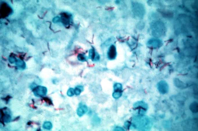
37 KB | Peter Anderson | This is a high-power (oil immersion) photomicrograph of granuloma stained with an acid-fast stain. Mycobacterium tuberculosis bacilli stain red. | 1 |
| 20:13, 20 August 2013 | IPLab6TB5.jpg (file) |  |
194 KB | Peter Anderson | High-power photomicrograph of a TB granuloma with multinucleated giant cells adjacent to an area of caseous necrosis (to the left). | 1 |
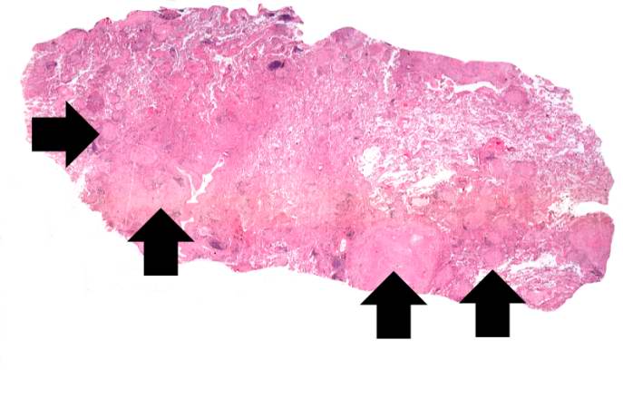
| 20:11, 20 August 2013 | IPLab6TB4.jpg (file) |  |
68 KB | Peter Anderson | This is a higher-power photomicrograph of a TB granuloma. The area of caseous necrosis is on the left side of the image, there are multinucleated giant cells and epithelioid macrophages throughout the remainder of the tissue. | 1 |
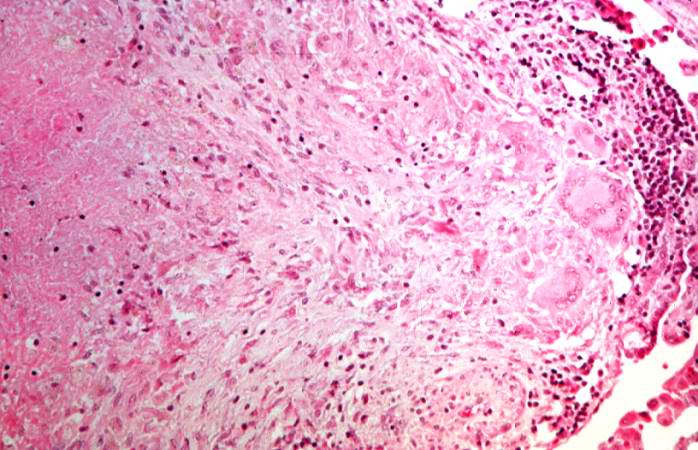
| 20:10, 20 August 2013 | IPLab6TB3.jpg (file) |  |
72 KB | Peter Anderson | This is a higher-power photomicrograph of a TB granuloma. Note the eosinophilic material in the center of this granuloma (caseous necrosis) and the epithelioid macrophages and giant cells around the periphery. | 1 |
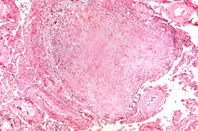
| 20:10, 20 August 2013 | IPLab6TB2.jpg (file) |  |
36 KB | Peter Anderson | This is a low-power photomicrograph of lung tissue with multiple circumscribed nodules - granulomas (arrows). | 1 |
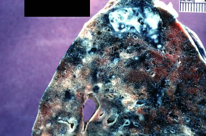
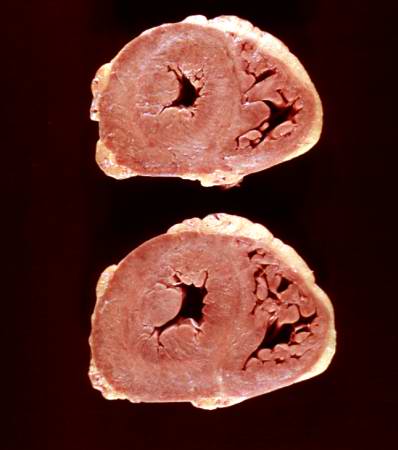
| 20:10, 20 August 2013 | IPLab6TB1.jpg (file) |  |
63 KB | Peter Anderson | This is a photograph of a section of lung with an apical lesion. This lesion represents an old healed lesion from Mycobacterium tuberculosis infection. | 1 |
| 19:59, 20 August 2013 | IPLab6Scleroderma5.jpg (file) |  |
19 KB | Peter Anderson | This is a gross photograph of the heart from this case. There is thickening of the left ventricular wall and some thickening of the right ventricle as well. | 1 |
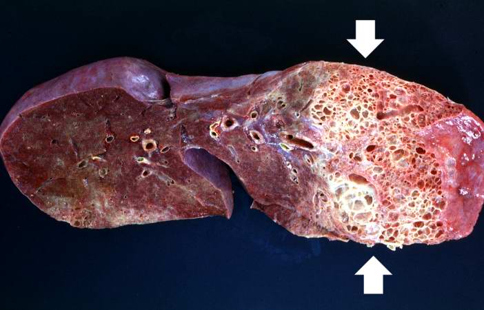
| 19:59, 20 August 2013 | IPLab6Scleroderma4.jpg (file) |  |
65 KB | Peter Anderson | This is a closer view of the cut section of lung from this patient showing the extensive fibrosis and the severe emphysematous change. | 1 |
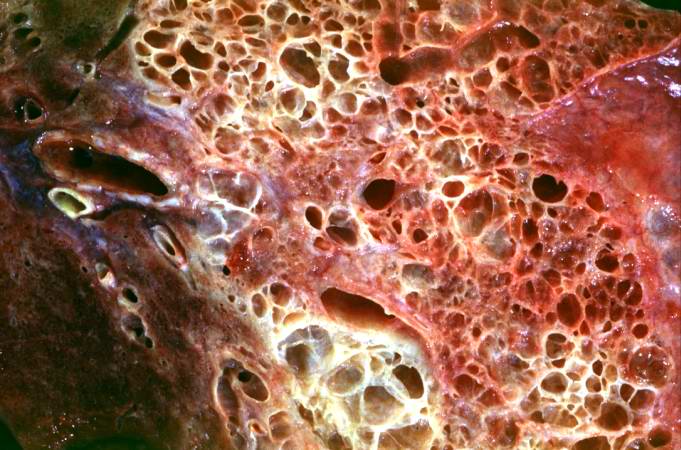
| 19:58, 20 August 2013 | IPLab6Scleroderma3.jpg (file) |  |
64 KB | Peter Anderson | This is a closer view of the cut section of lung from this patient. Note the extensive fibrosis and the severe emphysematous changes. | 1 |
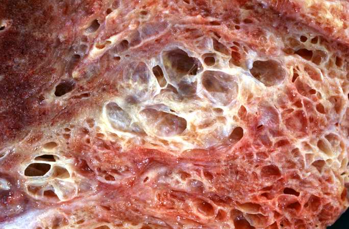
| 19:58, 20 August 2013 | IPLab6Scleroderma2.jpg (file) |  |
43 KB | Peter Anderson | This is a gross photograph of a cut section of one lung from this patient. Note the extensive fibrosis lower lobe (arrows). | 1 |
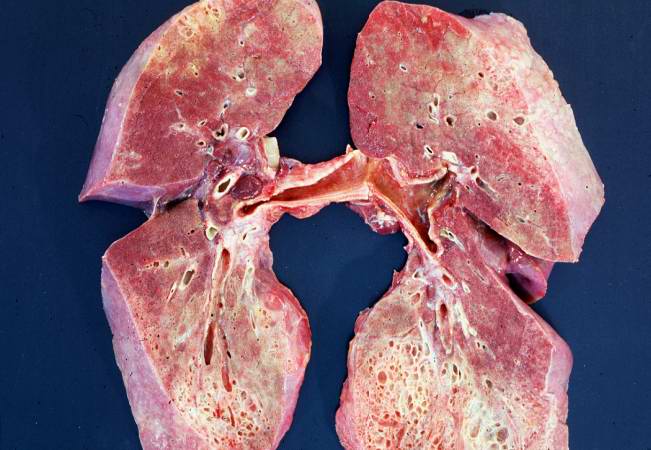
| 19:57, 20 August 2013 | IPLab6Scleroderma1.jpg (file) |  |
49 KB | Peter Anderson | This is a gross photograph of cut section of the lungs from this patient. Note the extensive fibrosis of the lung parenchyma. | 1 |
| 18:00, 20 August 2013 | IPLab6PAN13.jpg (file) |  |
85 KB | Peter Anderson | This is a high-power photomicrograph of the affected vessel in the heart. The vessel lumen is completely occluded. | 1 |
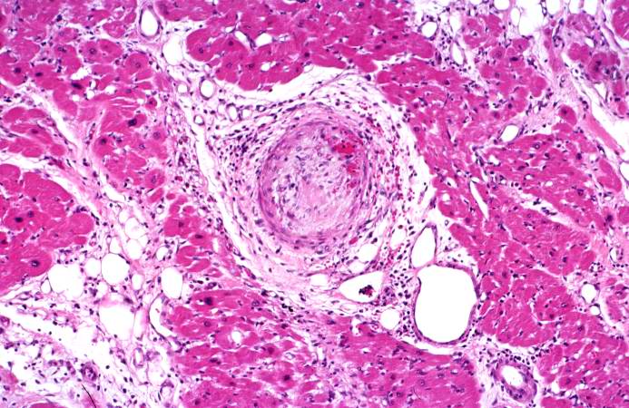
| 18:00, 20 August 2013 | IPLab6PAN12.jpg (file) |  |
88 KB | Peter Anderson | This is a higher-power photomicrograph of the affected vessels in the heart (arrows). There are areas of fibrosis (old infarcts) in the myocardium adjacent to these affected vessels. | 1 |
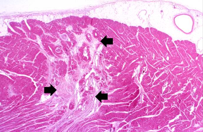
| 17:59, 20 August 2013 | IPLab6PAN11.jpg (file) |  |
69 KB | Peter Anderson | This is a low-power photomicrograph of the heart. There are areas of fibrosis in the myocardium (arrows). Note that the large epicardial coronary artery is normal. | 1 |
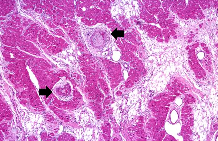
| 17:59, 20 August 2013 | IPLab6PAN10.jpg (file) |  |
76 KB | Peter Anderson | This is a higher-power photomicrograph of the affected vessel from the previous image. The vessel wall is infiltrated with inflammatory cells and the vessel lumen is completely occluded (arrow). | 1 |