File list
This special page shows all uploaded files.
| Date | Name | Thumbnail | Size | Description | Versions |
|---|---|---|---|---|---|
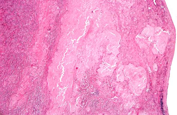
| 21:48, 20 August 2013 | IPLab6ChronicRejection1.jpg (file) |  |
43 KB | This is a low-power photomicrograph of the kidney from this case of chronic transplant rejection. Note the focal areas of hemorrhage and inflammatory cell infiltrate in this section. | 1 |
| 21:43, 20 August 2013 | IPLab6MM4.jpg (file) |  |
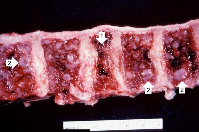
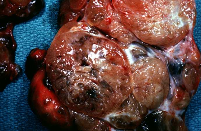
47 KB | This is a photograph of the vertebral column from this patient at autopsy. Notice the collapsed vertebra (1). There are multiple variably-sized white nodules (2) within the bone marrow. These are accumulations of malignant plasma cells in this case of ... | 1 |
| 21:43, 20 August 2013 | IPLab6MM3.jpg (file) |  |
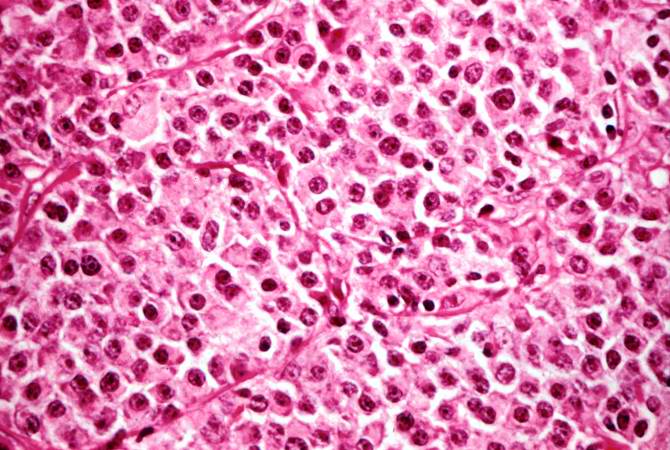
72 KB | This high-power photomicrograph demonstrates the cells that make up this tissue. These cells resemble plasma cells and are the malignant cell of multiple myeloma. | 1 |
| 21:42, 20 August 2013 | IPLab6MM2.jpg (file) |  |
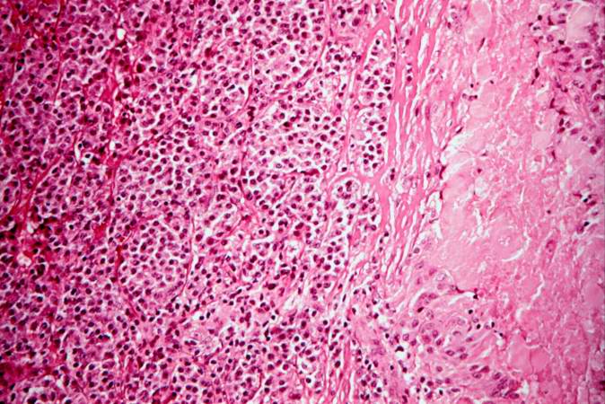
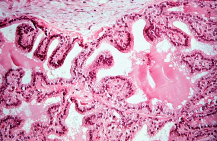
81 KB | This higher-power photomicrograph shows the junction between an amorphous hyaline-appearing area (amyloid) on the right and cellular areas (plasmacytoid cells) on the left. | 1 |
| 21:42, 20 August 2013 | IPLab6MM1.jpg (file) |  |
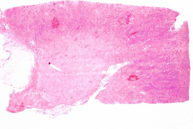
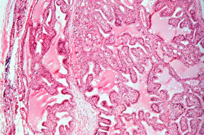
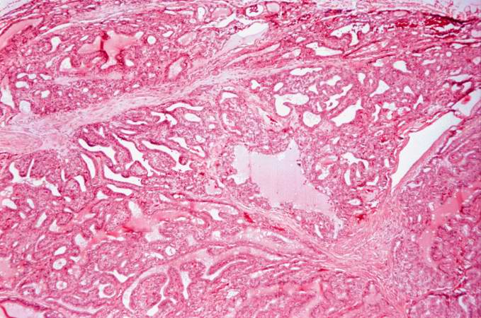
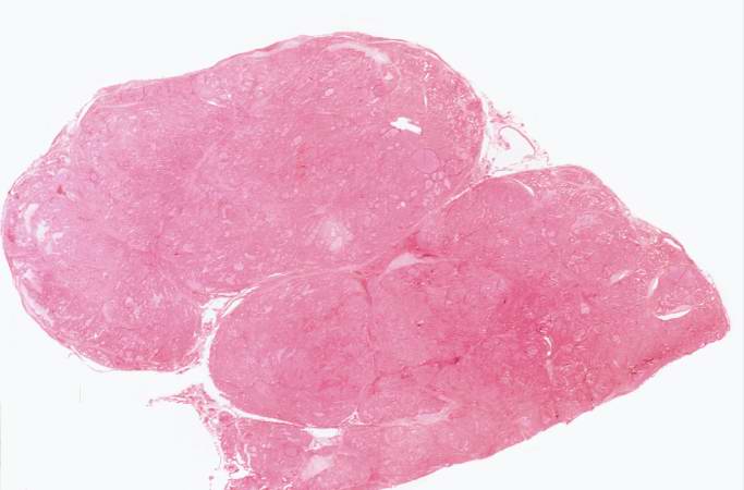
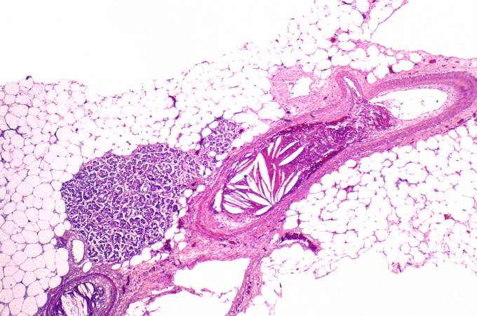
62 KB | This is a low-power photomicrograph of the mediastinal mass. The mass is encapsulated and contains cellular areas (blue) and areas of pale red material. | 1 |
| 21:36, 20 August 2013 | IPLab6Amyloid11.jpg (file) |  |
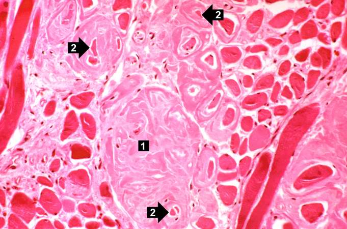
58 KB | This photomicrograph of the tongue demonstrates extensive amyloid deposits (1) separating the skeletal muscle fibers of the tongue. In many cases the amyloid encircles the muscle fibers (2) and these muscle fibers are atrophied. | 1 |
| 21:36, 20 August 2013 | IPLab6Amyloid10.jpg (file) |  |
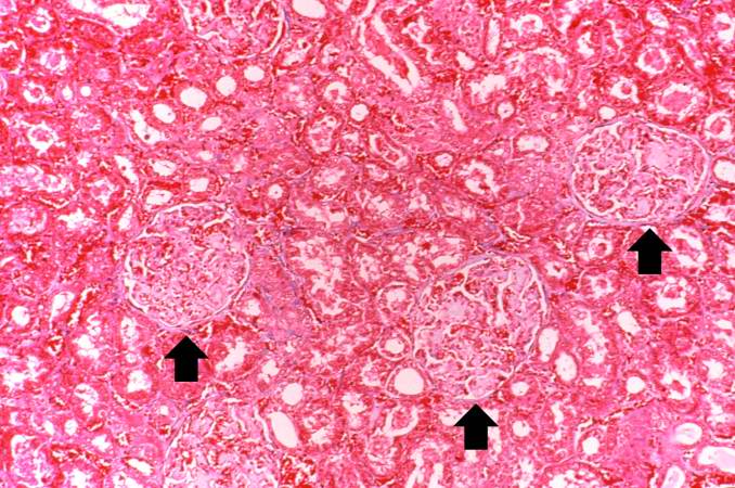
85 KB | This photomicrograph of kidney demonstrates the amyloid deposits (arrows) within glomeruli. | 1 |
| 21:36, 20 August 2013 | IPLab6Amyloid9.jpg (file) |  |
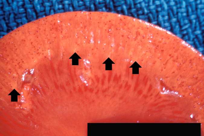
34 KB | This is a gross photograph of kidney from this case. Note the pale yellow material within the cortex (arrows). This is indicative of amyloid within the cortex and the glomeruli. Also note that there are multiple red spots in the cortex. These represent... | 1 |
| 21:35, 20 August 2013 | IPLab6Amyloid8.jpg (file) |  |
78 KB | This is a photomicrograph of Congo-red-stained liver tissue viewed with partially polarized light. Although not well demonstrated in this image, Congo-red-stained amyloid viewed through polarized light should give off a classic “apple green” birefr... | 1 |
| 21:35, 20 August 2013 | IPLab6Amyloid7.jpg (file) |  |
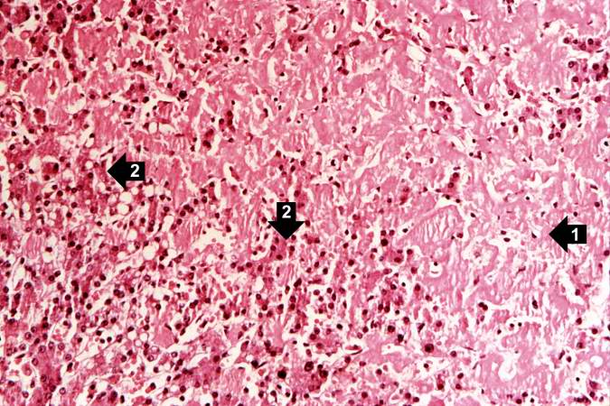
75 KB | This is a high-power view of liver tissue stained with Congo red. The orange amyloid material (arrows) is seen clearly between liver parenchymal cells. | 1 |
| 21:35, 20 August 2013 | IPLab6Amyloid6.jpg (file) |  |
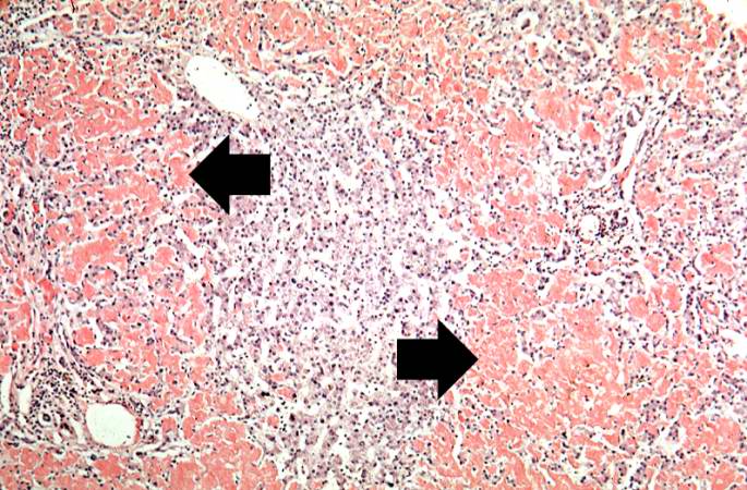
92 KB | This is a low-power photomicrograph of liver tissue stained with Congo red (orange color in slide). Congo red reacts with amyloid and gives it an orange color (arrows). | 1 |
| 21:34, 20 August 2013 | IPLab6Amyloid5.jpg (file) |  |
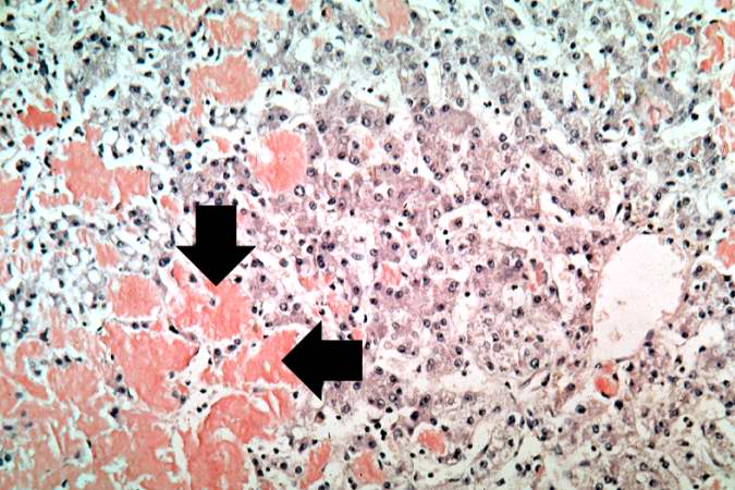
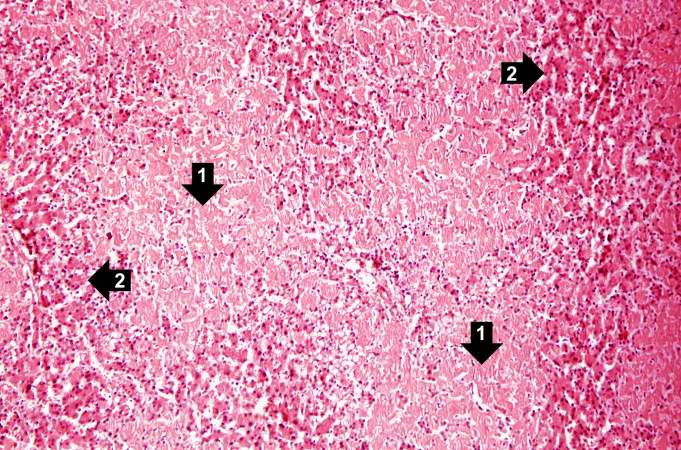
85 KB | This is a higher-power photomicrograph showing the amyloid deposits (1) between hepatocytes (2). | 1 |
| 21:34, 20 August 2013 | IPLab6Amyloid4.jpg (file) |  |
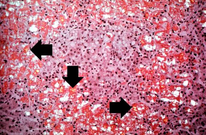
97 KB | This is a low-power photomicrograph of liver tissue from this case. Note the eosinophilic hyaline material (1) present within and between the hepatic tissue (2). There is marked distortion of lobular architecture by the amyloid. | 1 |
| 21:34, 20 August 2013 | IPLab6Amyloid3.jpg (file) |  |
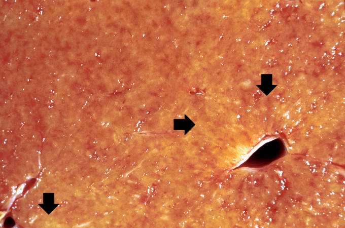
47 KB | This is a closer view of the cut surface of this liver. The pale waxy material can be seen within the hepatic tissue (arrows). | 1 |
| 21:33, 20 August 2013 | IPLab6Amyloid2.jpg (file) |  |
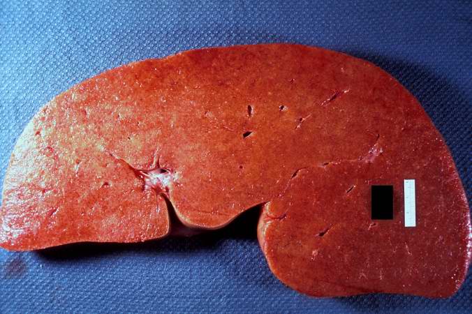
60 KB | This is a gross picture of the cut surface of the liver from this case. The liver tissue is firm and has a waxy appearance--although this is difficult to appreciate in an image. | 1 |
| 21:33, 20 August 2013 | IPLab6Amyloid1.jpg (file) |  |
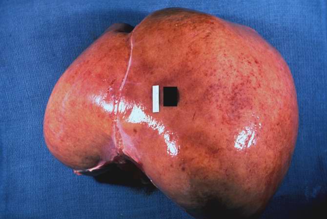
35 KB | This is a gross picture of liver from this case. Note the pale, swollen appearance of this liver. | 1 |
| 20:25, 20 August 2013 | IPLab6SenileAmyloidosis5.jpg (file) |  |
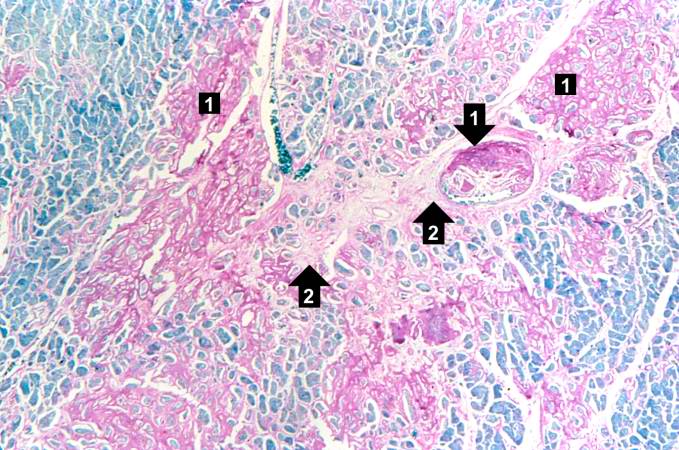
80 KB | This is a special stain for amyloid (Luxol PAS) demonstrating the amyloid (1) and fibrosis (2) in the myocardium. The amyloid is darker purple/magenta and tends to be more amorphous. The fibrosis is pink and more fibrillar. | 1 |
| 20:25, 20 August 2013 | IPLab6SenileAmyloidosis4.jpg (file) |  |
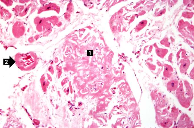
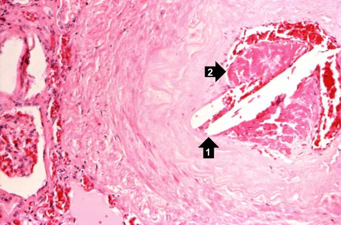
53 KB | This is a higher-power photomicrograph of extracellular amyloid (1) and deposition of amyloid in the vessel wall (2). | 1 |
| 20:24, 20 August 2013 | IPLab6SenileAmyloidosis3.jpg (file) |  |
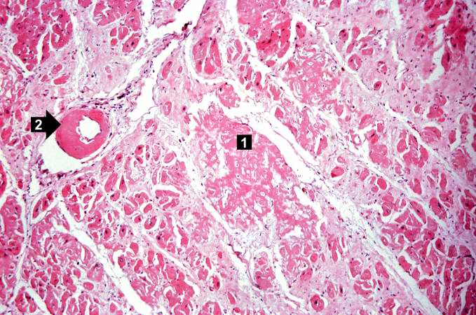
81 KB | This is a higher-power photomicrograph of the heart tissue from this case. Note the amyloid deposition throughout the myocardium (1) as well as deposition in the wall of the blood vessel (2). | 1 |
| 20:24, 20 August 2013 | IPLab6SenileAmyloidosis2.jpg (file) |  |
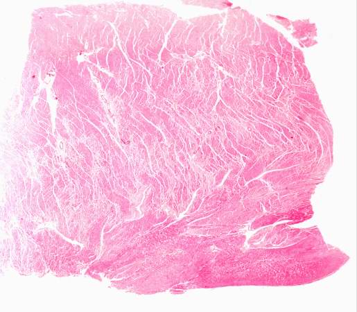
39 KB | This is a low power photomicrograph of the heart tissue from this case. At this magnification the structure looks relatively normal. | 1 |
| 20:24, 20 August 2013 | IPLab6SenileAmyloidosis1.jpg (file) |  |
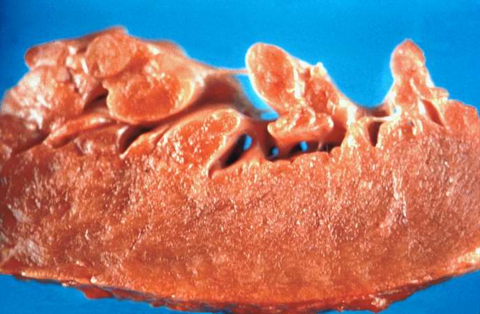
47 KB | This is a gross photograph of section of heart tissue from this case. The tissue was firm and had a waxy texture. If you use your imagination you can see pale yellow areas within this tissue which represent the amyloid deposits. | 1 |
| 18:29, 19 August 2013 | IPLab5Downs6.jpg (file) |  |
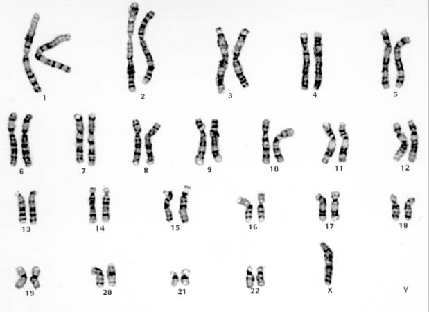
20 KB | This is a karyotype of a patient with Turner syndrome (45, X). | 1 |
| 18:28, 19 August 2013 | IPLab5Downs5.jpg (file) |  |
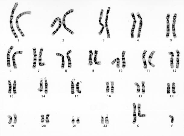
19 KB | This is a karyotype of a patient with Klinefelter syndrome (47, XXY). | 1 |
| 18:28, 19 August 2013 | IPLab5Downs4.jpg (file) |  |
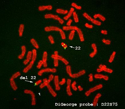
22 KB | FISH is also useful in the diagnosis of other genetic disorders. This is an example of FISH staining on another patient using a probe specific for DiGeorge's disease. The arrow shows that there is a deletion on chromosome 22, which is diagnostic for Di... | 1 |
| 18:28, 19 August 2013 | IPLab5Downs3.jpg (file) |  |
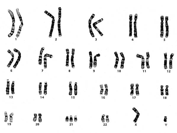
21 KB | Chromosomes from the chromosome spread are lined up to demonstrate the karyotype. In this case there are three copies of chromosome 21, just as noted in the FISH. | 1 |
| 18:27, 19 August 2013 | IPLab5Downs2.jpg (file) |  |
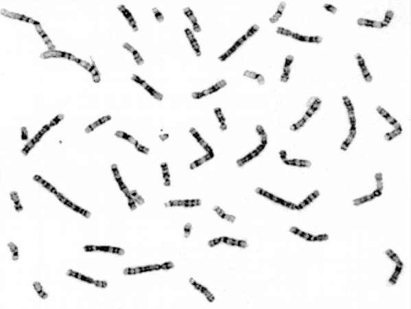
17 KB | These cells, obtained by amniocentesis, were cultured and then arrested in metaphase. Nuclei from these cells were isolated and stained to demonstrate the banding pattern of each chromosome. This photograph shows a "chromosome spread." Each chromosome ... | 1 |
| 18:27, 19 August 2013 | IPLab5Downs1.jpg (file) |  |
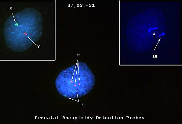
18 KB | This is a photomicrograph of cells obtained by amniocentesis that were stained using FISH. The cell in panel 1 was stained with markers specific for the X and Y-chromosomes. The cell in panel 2 was stained with a marker specific for chromosome 18. The ... | 1 |
| 18:23, 19 August 2013 | IPLab6GravesDisease7.jpg (file) |  |
61 KB | Closer view of cut surface of the thyroid with nodular goiter. Note the multilobular appearance of the tissue. | 1 |
| 18:23, 19 August 2013 | IPLab6GravesDisease6.jpg (file) |  |
63 KB | This is a gross photograph of a thyroid from a case of nodular goiter. | 1 |
| 18:23, 19 August 2013 | IPLab6GravesDisease5.jpg (file) |  |
65 KB | This is a high-power photomicrograph of thyroid. Note the papillary projections and the moth-eaten appearance of the colloid. This appearance indicates active absorption of the colloid to form thyroglobulin. | 1 |
| 18:22, 19 August 2013 | IPLab6GravesDisease4.jpg (file) |  |
77 KB | This is a high-power photomicrograph of thyroid. Note the cellularity of the tissue with marked infolding of the epithelial tissue. | 1 |
| 18:22, 19 August 2013 | IPLab6GravesDisease3.jpg (file) |  |
75 KB | This is a higher-power photomicrograph of thyroid. The tissue is very cellular and there is little colloid. | 1 |
| 18:22, 19 August 2013 | IPLab6GravesDisease2.jpg (file) |  |
29 KB | This is a low-power photomicrograph of thyroid tissue from this case. The tissue is very cellular with very little colloid. | 1 |
| 18:21, 19 August 2013 | IPLab6GravesDisease1.jpg (file) |  |
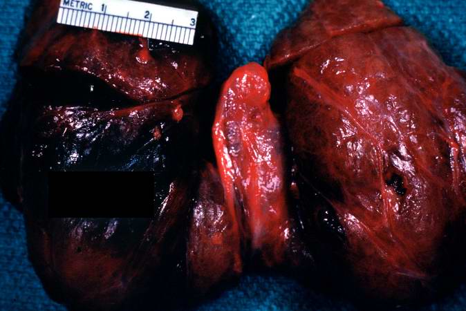
43 KB | This is a photograph of the thyroid from this case. Note that the gland is enlarged and is dark red. | 1 |
| 18:09, 19 August 2013 | IPLab6RA10.jpg (file) |  |
65 KB | This is a high-power photomicrograph of another region with macrophages (right), fibrocytes (left), and occasional lymphocytes throughout the lesion. | 1 |
| 18:08, 19 August 2013 | IPLab6RA9.jpg (file) |  |
63 KB | This is a high-power photomicrograph of the mononuclear cells which surround the central area of necrosis. The focal accumulations of fibrinoid material are clearly visible. Lymphocytes are present in the extreme right of this image. | 1 |
| 18:08, 19 August 2013 | IPLab6RA8.jpg (file) |  |
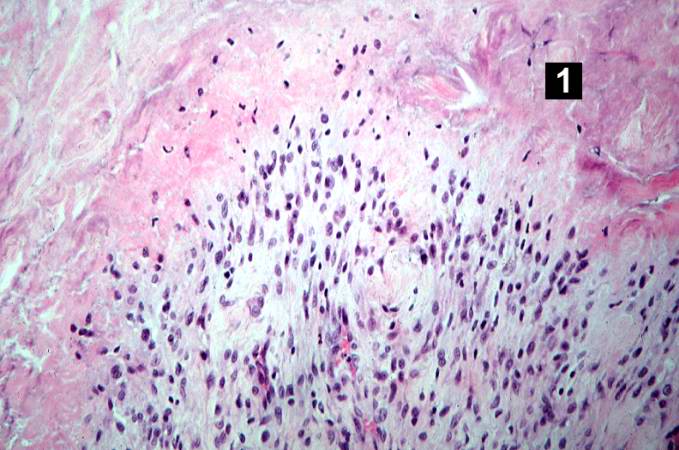
59 KB | This higher-power photomicrograph of the tissue illustrates the palisading nuclei of the monocytes which are located around the periphery of the central necrotic region (1). | 1 |
| 18:07, 19 August 2013 | IPLab6RA7.jpg (file) |  |
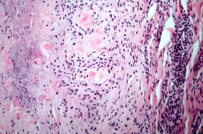
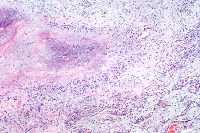
82 KB | This higher-power photomicrograph of the subcutaneous nodule again demonstrates the necrotic center and peripheral rim of macrophages, fibrocytes, and occasional lymphocytes. There are focal accumulations of hyaline material (fibrinoid material) within... | 1 |
| 18:07, 19 August 2013 | IPLab6RA6.jpg (file) |  |
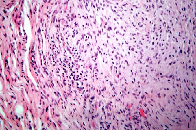
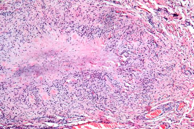
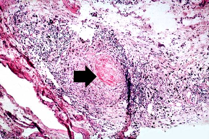
64 KB | This higher-power photomicrograph of the subcutaneous nodule shows a granulomatous lesion with a necrotic center and a peripheral rim of macrophages, fibrocytes, and occasional lymphocytes. In the necrotic center of the granuloma there is some minerali... | 1 |
| 18:07, 19 August 2013 | IPLab6RA5.jpg (file) |  |
11 KB | This is a low-power photomicrograph of the subcutaneous nodule from this patient. | 1 |
| 18:06, 19 August 2013 | IPLab6RA4.jpg (file) |  |
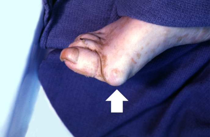
24 KB | This is a gross photograph of the foot from this same patient. Note the subcutaneous nodule on the medial aspect of the foot (arrow). | 1 |
| 18:06, 19 August 2013 | IPLab6RA3.jpg (file) |  |
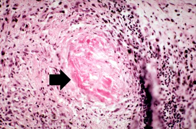
62 KB | This is a high-power photomicrograph of the joint capsule with another granuloma surrounding a central area of fibrinoid necrosis (arrow). | 1 |
| 18:06, 19 August 2013 | IPLab6RA2.jpg (file) |  |
97 KB | This is a medium-power photomicrograph of the joint capsule surrounding the metacarpal joints. Note the thickening of the capsule and the focal accumulation of inflammatory cells surrounding a central area of fibrinoid necrosis (arrow). | 1 |
| 18:05, 19 August 2013 | IPLab6RA1.jpg (file) |  |
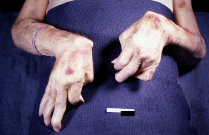
34 KB | This is a gross photograph of the patient's hands at autopsy. Note the swollen joints and the deforming arthritis. | 1 |
| 17:01, 19 August 2013 | IPLab4AtheromatousEmboli6.jpg (file) |  |
62 KB | A mesenteric artery also had an atherosclerotic embolus. Again note the cholesterol clefts and thrombotic material that occlude this artery. | 1 |
| 17:01, 19 August 2013 | IPLab4AtheromatousEmboli5.jpg (file) |  |
54 KB | This is another view of this vessel with an atherosclerotic embolus. Note the cholesterol clefts (1) and thrombotic material (2) that occlude this artery. | 1 |
| 17:01, 19 August 2013 | IPLab4AtheromatousEmboli4.jpg (file) |  |
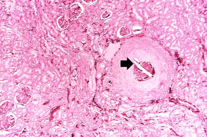
78 KB | This higher-power photomicrograph of one of the arcuate arteries reveals a cholesterol embolus. Note the needle-shaped space (arrow) within the lumen of this artery (arrow) which represents the space occupied by the cholesterol crystal that was dissolv... | 1 |
| 17:00, 19 August 2013 | IPLab4AtheromatousEmboli3.jpg (file) |  |
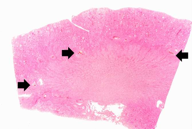
34 KB | This is a low-power photomicrograph of kidney tissue. Several blood vessels can be identified at the corticomedullary junction (arrows). | 1 |
| 17:00, 19 August 2013 | IPLab4AtheromatousEmboli2.jpg (file) |  |
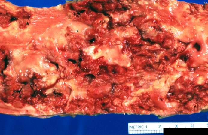
53 KB | This is a closer view of the luminal surface of the aorta from the previous image. The rough, ulcerated surface and the thrombotic material can be easily seen in this image. | 1 |
| 17:00, 19 August 2013 | IPLab4AtheromatousEmboli1.jpg (file) |  |
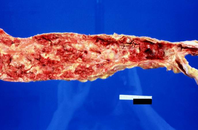
31 KB | This is a gross photograph of the aorta from this patient opened lengthwise with the luminal surface visible. Note the rough surface with ulcerations and adherent thrombotic material. There is a mild dilation (aneurysm) at the distal aorta just at the ... | 1 |