File list
This special page shows all uploaded files.
| Date | Name | Thumbnail | Size | User | Description | Versions |
|---|---|---|---|---|---|---|
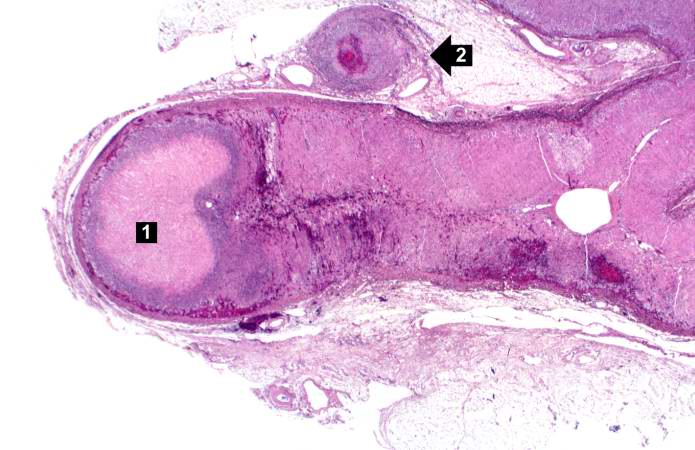
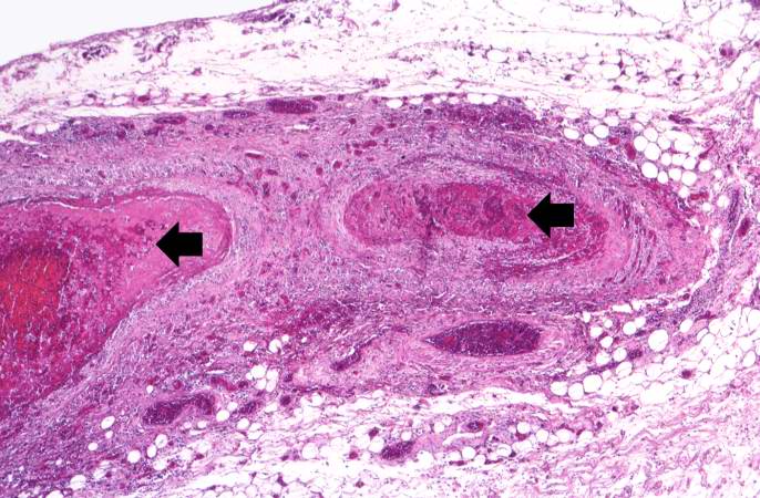
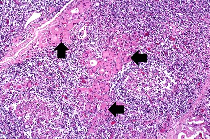
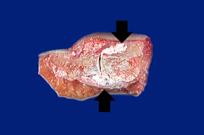
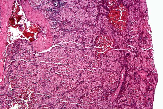
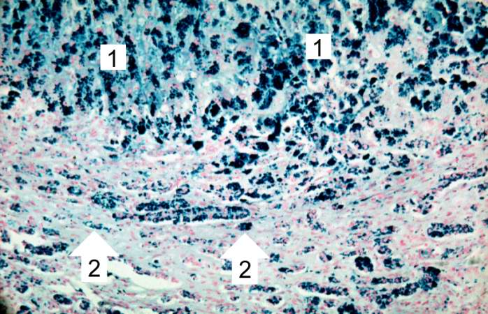
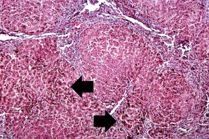
| 17:58, 20 August 2013 | IPLab6PAN9.jpg (file) |  |
53 KB | Peter Anderson | This is a low-power photomicrograph of the adrenal gland. There is an area of necrosis in the adrenal (1) and an affected vessel adjacent to the adrenal (2). | 1 |
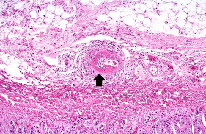
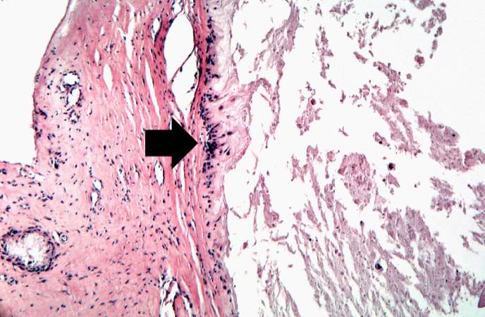
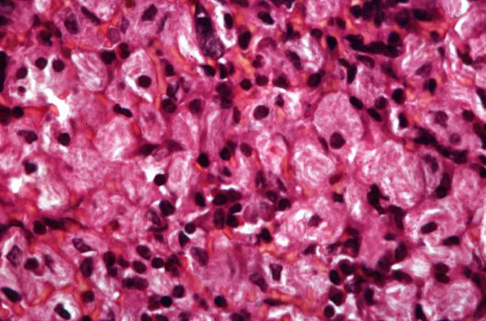
| 17:58, 20 August 2013 | IPLab6PAN8.jpg (file) |  |
86 KB | Peter Anderson | This is a high-power photomicrograph of a small vessel with a rim of fibrinoid necrosis (arrow). | 1 |
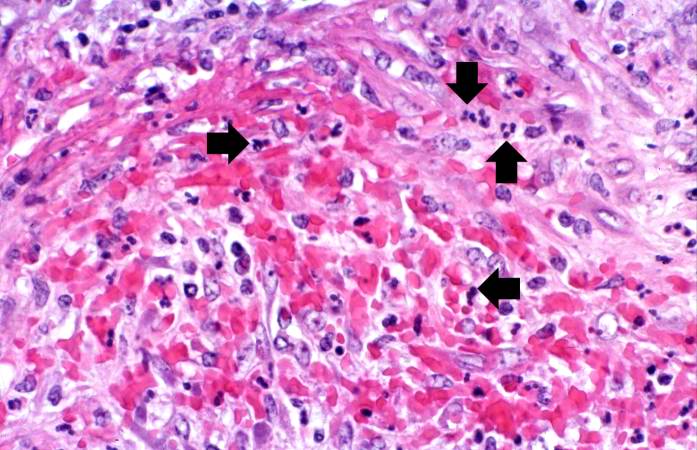
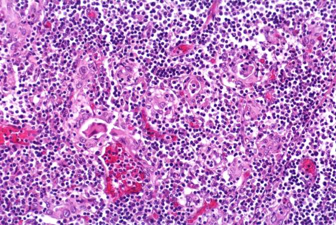
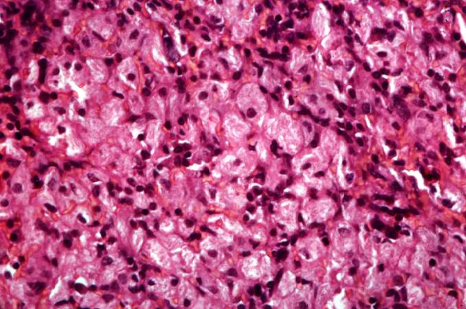
| 17:57, 20 August 2013 | IPLab6PAN7.jpg (file) |  |
67 KB | Peter Anderson | This is a high-power photomicrograph of the vessel wall. There is hemorrhage and infiltration with inflammatory cells--primarily neutrophils (arrows). | 1 |
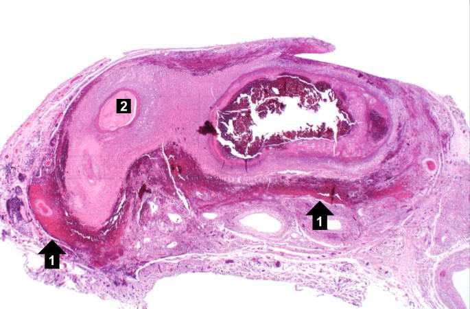
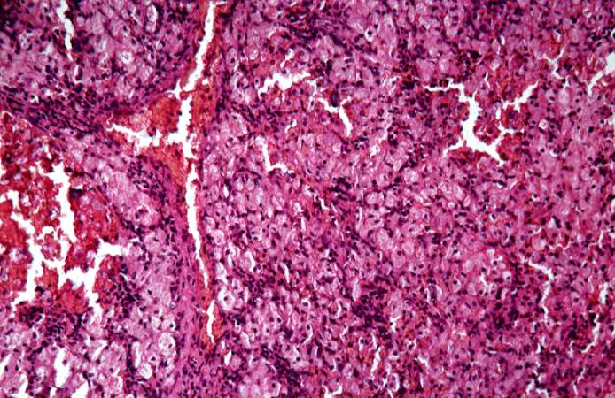
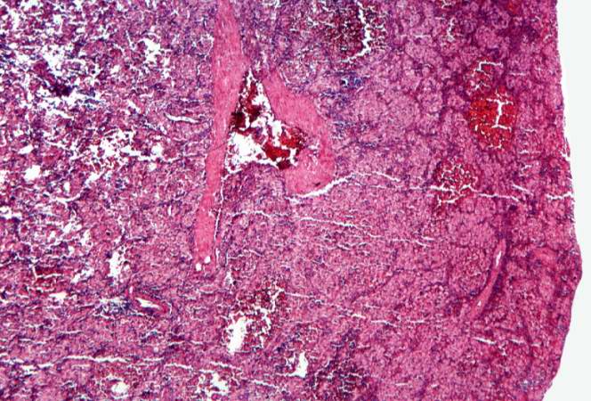
| 17:57, 20 August 2013 | IPLab6PAN6.jpg (file) |  |
50 KB | Peter Anderson | his is another example of a mesenteric artery from this case. There is a marked inflammatory cell response surrounding this vessel, fresh hemorrhage (1), and thrombotic material (2). | 1 |
| 17:56, 20 August 2013 | IPLab6PAN5.jpg (file) |  |
79 KB | Peter Anderson | This is a higher-power photomicrograph of this mesenteric vessel. Note the thrombotic material occluding the vessel (arrows) and the inflammatory cell infiltrate in the wall of the vessel and in the surrounding adventitia. | 1 |
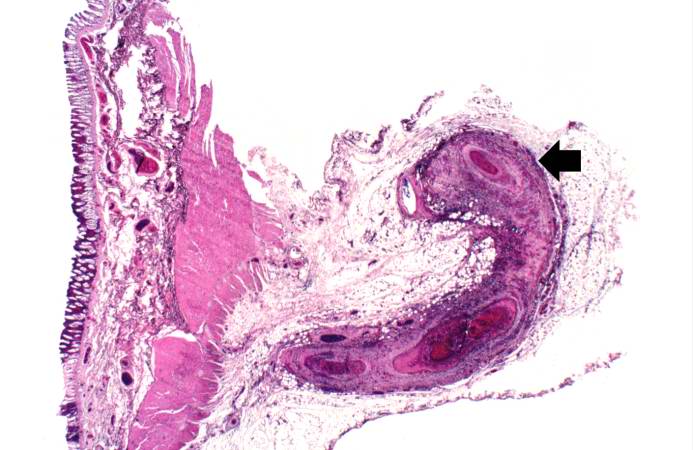
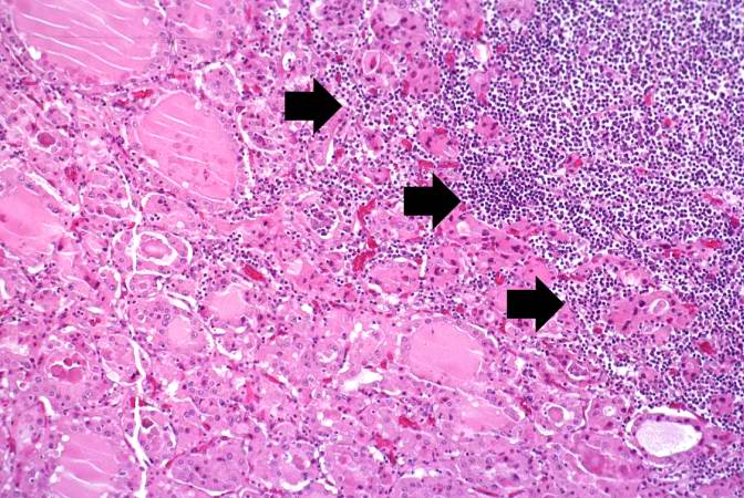
| 17:56, 20 August 2013 | IPLab6PAN4.jpg (file) |  |
51 KB | Peter Anderson | This is a low-power photomicrograph of a mesenteric vessel from this case of polyarteritis nodosa (arrow). The vessel is completely occluded by thrombotic material and the vessel wall is infiltrated with inflammatory cells. | 1 |
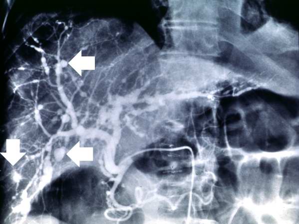
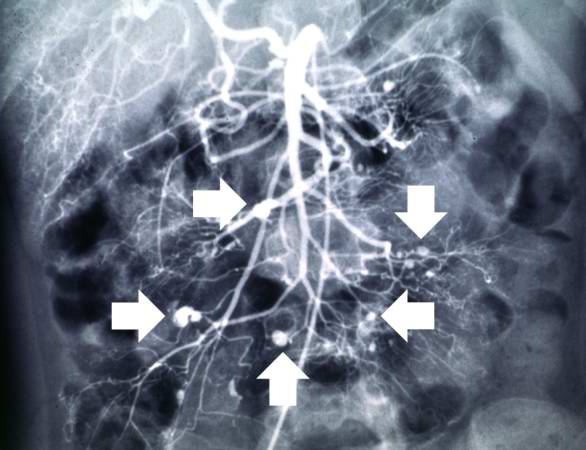
| 17:56, 20 August 2013 | IPLab6PAN3.jpg (file) |  |
36 KB | Peter Anderson | This angiogram of the kidneys demonstrates numerous aneurysmal dilatations in the renal circulation (arrows). | 1 |
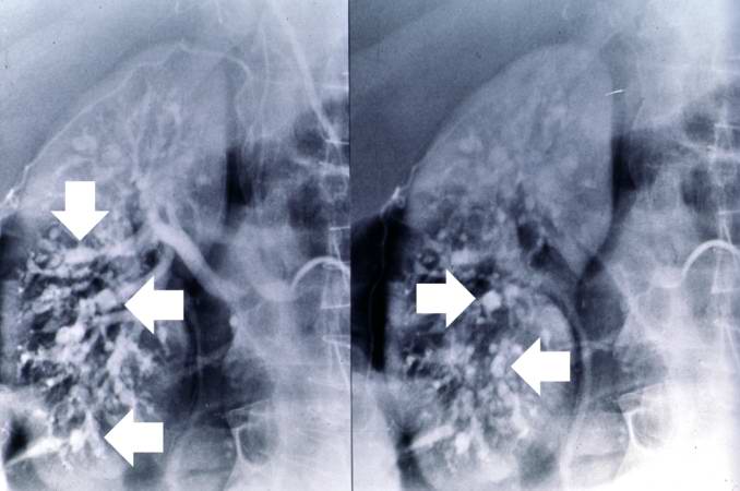
| 17:55, 20 August 2013 | IPLab6PAN2.jpg (file) |  |
36 KB | Peter Anderson | This angiogram of the liver also demonstrates numerous aneurysms throughout the hepatic circulation (arrows). | 1 |
| 17:55, 20 August 2013 | IPLab6PAN1.jpg (file) |  |
36 KB | Peter Anderson | This angiogram of the abdominal viscera demonstrates numerous aneurysms throughout the mesenteric circulation (arrows). | 1 |
| 17:47, 20 August 2013 | IPLab6Hashimoto9.jpg (file) | 111 KB | Peter Anderson | This high-power photomicrograph shows more clearly the lymphocytes and plasma cells surrounding the thyroid gland epithelium. Large, eosinophilic, degenerating thyroid gland cells (Hurthle cells) can be seen in this section (arrows). | 1 | |
| 17:47, 20 August 2013 | IPLab6Hashimoto8.jpg (file) | 94 KB | Peter Anderson | This is a high-power photomicrograph showing the lymphocytes and plasma cells surrounding the thyroid gland epithelium. | 1 | |
| 17:46, 20 August 2013 | IPLab6Hashimoto7.jpg (file) | 87 KB | Peter Anderson | This is a high-power photomicrograph showing the inflammatory cells infiltrating into the residual thyroid tissue (arrows). | 1 | |
| 17:46, 20 August 2013 | IPLab6Hashimoto6.jpg (file) | 75 KB | Peter Anderson | This is another higher-power photomicrograph of thyroid from this case showing the inflammatory cells and the residual thyroid tissue. | 1 | |
| 17:44, 20 August 2013 | IPLab6Hashimoto5.jpg (file) | 76 KB | Peter Anderson | This is a higher-power photomicrograph of thyroid from this case showing the inflammatory cells and the residual thyroid tissue. | 1 | |
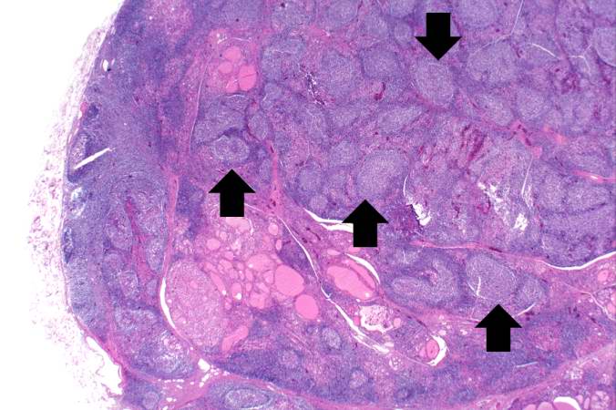
| 17:43, 20 August 2013 | IPLab6Hashimoto4.jpg (file) | 58 KB | Peter Anderson | This is another view of thyroid gland filled with inflammatory cells forming germinal centers (arrows). | 1 | |
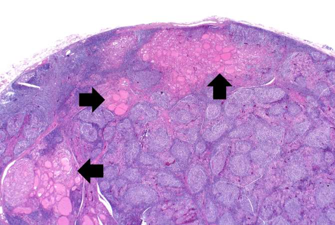
| 17:43, 20 August 2013 | IPLab6Hashimoto3.jpg (file) | 55 KB | Peter Anderson | This is a higher-power photomicrograph of thyroid from this case. Note the large number of blue-staining inflammatory cells in this tissue. These cells appear to be forming germinal centers. Some residual thyroid gland tissue can be seen in this sectio... | 1 | |
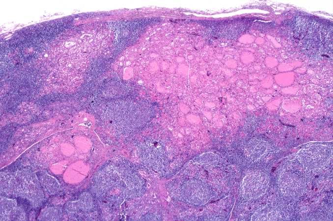
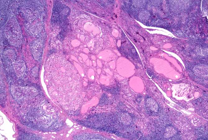
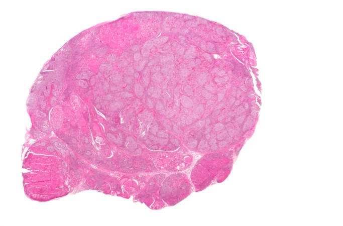
| 17:42, 20 August 2013 | IPLab6Hashimoto2.jpg (file) | 27 KB | Peter Anderson | This is a low-power photomicrograph of thyroid from this case. Note that the tissue is more cellular than one would expect and there does not appear to be normal colloid-filled blue spaces in this gland. | 1 | |
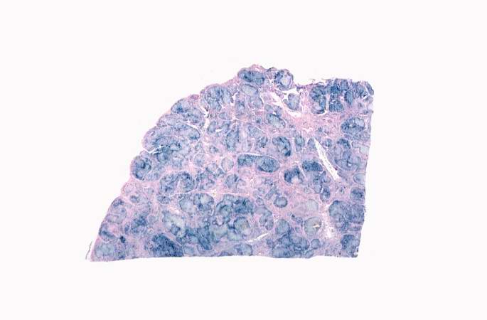
| 17:42, 20 August 2013 | IPLab6Hashimoto1.jpg (file) | 20 KB | Peter Anderson | This is a gross photograph of thyroid gland taken at autopsy. The gland is only slightly enlarged and has a firm texture. | 1 | |
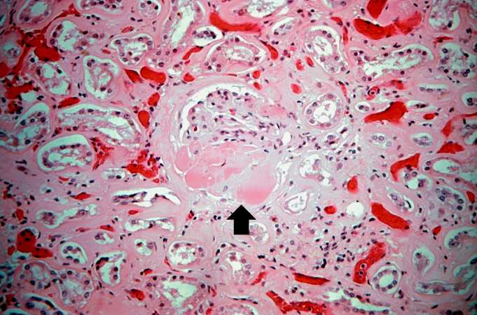
| 15:29, 20 August 2013 | IPLab5DM7.jpg (file) |  |
66 KB | Peter Anderson | This is a photomicrograph of kidney with a focal exudative lesion in a glomerulus (arrow) and sclerosis, interstitial fibrosis, and congestion. | 1 |
| 15:28, 20 August 2013 | IPLab5DM6.jpg (file) |  |
56 KB | Peter Anderson | This is a higher-power photomicrograph of a glomerulus with nodular glomerulosclerosis (arrows). These are the classic Kimmelstiel-Wilson lesions ("K-W lesions") seen in diabetics with nodular glomerulosclerosis. | 1 |
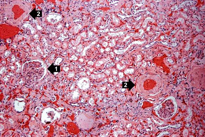
| 15:27, 20 August 2013 | IPLab5DM5.jpg (file) |  |
74 KB | Peter Anderson | This is a photomicrograph of a glomerulus with nodular glomerulosclerosis (1). Also note the intertubular fibrosis and the changes in the blood vessels (2). | 1 |
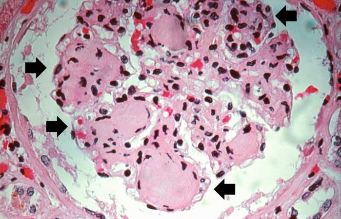
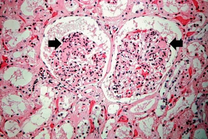
| 15:27, 20 August 2013 | IPLab5DM4.jpg (file) |  |
74 KB | Peter Anderson | This is a high-power photomicrograph of two glomeruli with intercapillary glomerulosclerosis (arrows). | 1 |
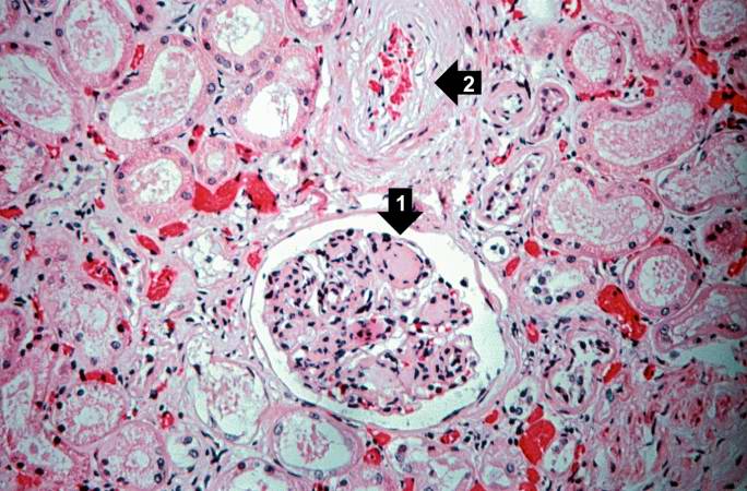
| 15:27, 20 August 2013 | IPLab5DM3.jpg (file) |  |
94 KB | Peter Anderson | This is a higher-power photomicrograph of the cortical region. In this region there is ischemic obsolescence of glomeruli and one glomerulus with nodular glomerulosclerosis (1). Also note the thickened walls of the blood vessels (2). | 1 |
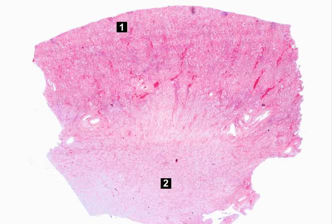
| 15:26, 20 August 2013 | IPLab5DM2.jpg (file) |  |
36 KB | Peter Anderson | This is a low-power photomicrograph of the kidney from this patient. The section extends from cortex (1) to the medulla (2). | 1 |
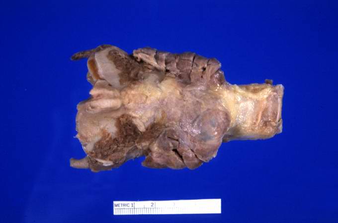
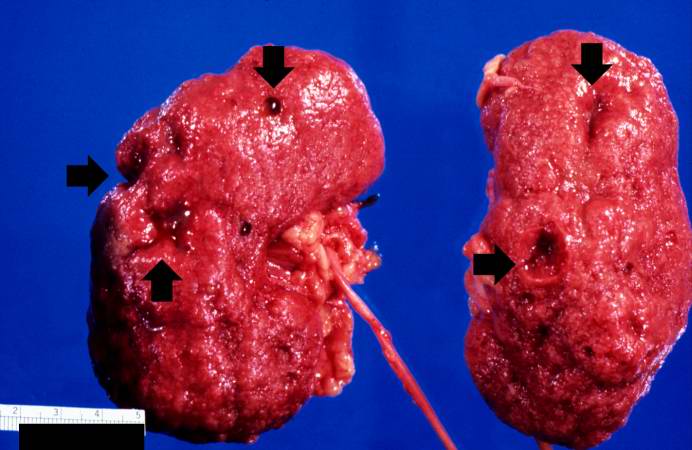
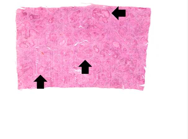
| 15:26, 20 August 2013 | IPLab5DM1.jpg (file) |  |
44 KB | Peter Anderson | This is a gross photograph of the kidneys from this case. Note that there are multiple shrunken regions (old infarcts) (arrows) and the kidneys have a rough granular appearance on the surface, which is caused by multiple small infarcts of small vessels... | 1 |
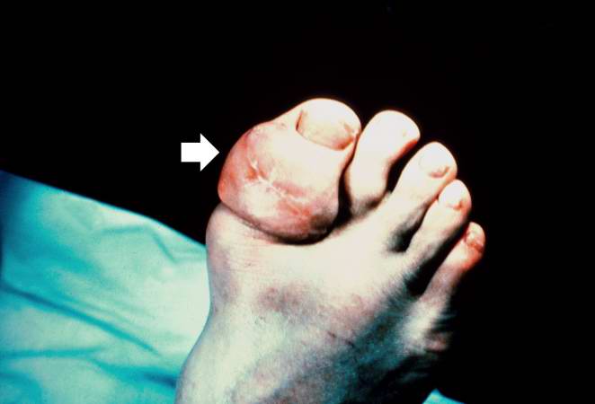
| 15:20, 20 August 2013 | IPLab5Gout8.jpg (file) |  |
20 KB | Peter Anderson | This is a gross photograph of a tophus on the great toe of another patient with gout (arrow). The healed surgical incision and the size of this tophus indicate that this was a long-standing problem for this patient. | 1 |
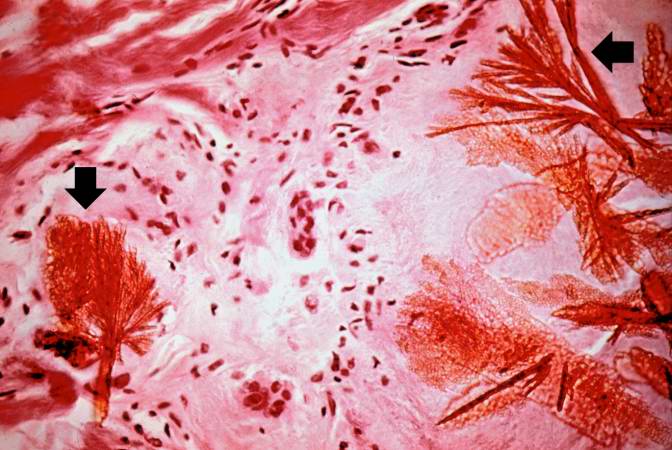
| 15:20, 20 August 2013 | IPLab5Gout7.jpg (file) |  |
59 KB | Peter Anderson | This is a photomicrograph of a tophus that was fixed in alcohol prior to histologic processing. The alcohol fixation preserves the water soluble urate crystals within the tissue. Note the urate crystals visible in this photomicrograph (arrows). Also no... | 1 |
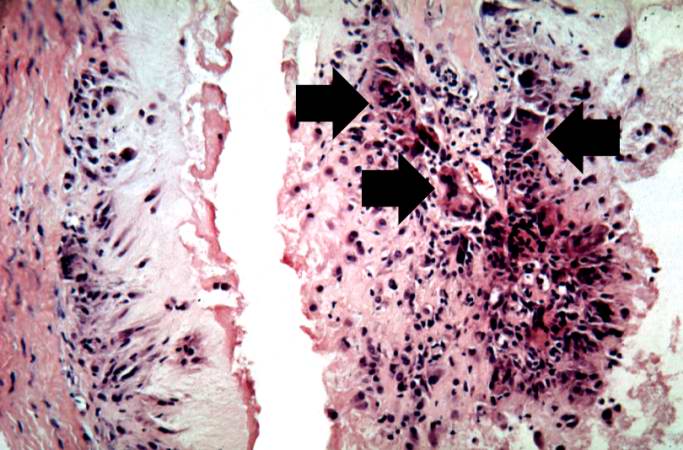
| 15:20, 20 August 2013 | IPLab5Gout6.jpg (file) |  |
63 KB | Peter Anderson | This is a high-power photomicrograph of the edge of the tophus. The character of the intense chronic inflammatory cell reaction is evident and note the presence of giant cells within this inflammatory cell reaction (arrows). | 1 |
| 15:19, 20 August 2013 | IPLab5Gout5.jpg (file) |  |
60 KB | Peter Anderson | This is a higher-power photomicrograph of the edge of the tophus. Most of the urate crystals dissolve away during processing. The inflammatory cells at the edge of these foci are clearly visible (arrow). | 1 |
| 15:19, 20 August 2013 | IPLab5Gout4.jpg (file) |  |
61 KB | Peter Anderson | This higher-power photomicrograph of the tophus demonstrates the collections of urate crystals (1) and the inflammatory cells at the edge of these foci (2). | 1 |
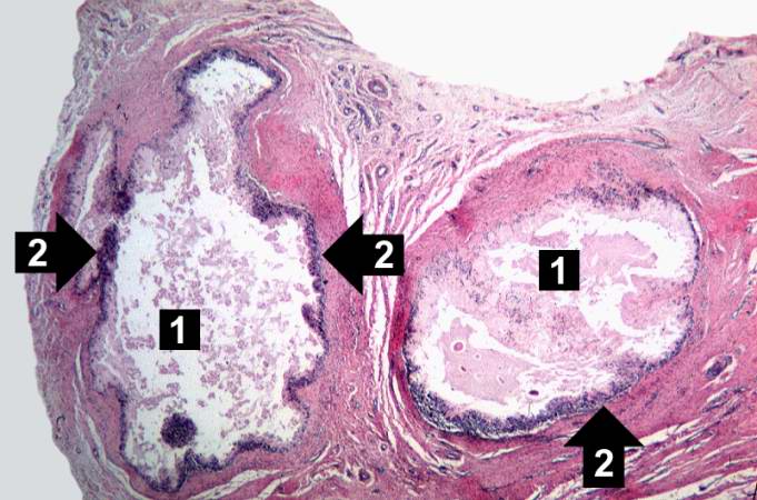
| 15:18, 20 August 2013 | IPLab5Gout3.jpg (file) |  |
21 KB | Peter Anderson | This is a low-power photomicrograph of the tophus removed from the elbow of this patient. Note the fibrous connective tissue (1) and the large foci containing the urate crystals (2) surrounded by the intense chronic inflammatory reaction. | 1 |
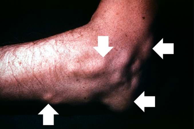
| 15:18, 20 August 2013 | IPLab5Gout2.jpg (file) |  |
26 KB | Peter Anderson | This is a gross photograph of the elbow of this patient. The subcutaneous nodules (arrows) on this arm are tophi caused by gout. | 1 |
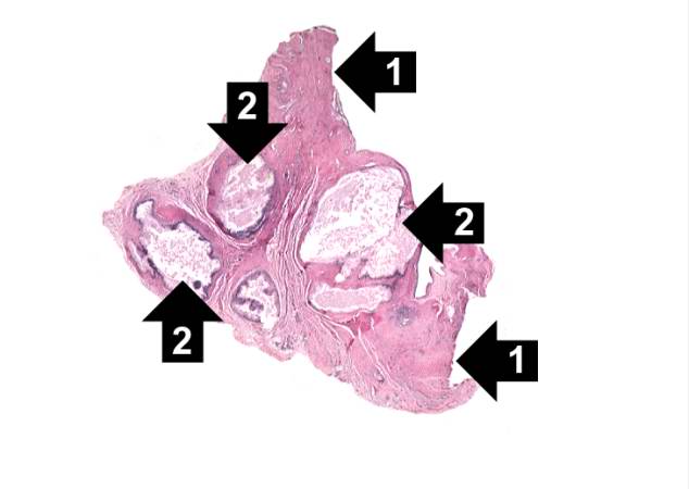
| 15:17, 20 August 2013 | IPLab5Gout1.jpg (file) |  |
22 KB | Peter Anderson | This is a gross photograph of an index finger from a patient with gout. The finger has been sectioned longitudinally to demonstrate the distal interphalangeal joint. Note the white chalky material within and adjacent to the joint (arrows). | 1 |
| 15:12, 20 August 2013 | IPLab5Gaucher8.jpg (file) |  |
53 KB | Peter Anderson | This is a higher-power photomicrograph of the spleen from this case. At this higher power individual cells can be better appreciated and the fibrillar nature of the eosinophilic cytoplasmic material can be seen. | 1 |
| 15:12, 20 August 2013 | IPLab5Gaucher7.jpg (file) |  |
64 KB | Peter Anderson | This is another high-power photomicrograph of the spleen from this case. At this high power individual cells can be better appreciated. | 1 |
| 15:11, 20 August 2013 | IPLab5Gaucher6.jpg (file) |  |
87 KB | Peter Anderson | This is another high-power photomicrograph of the spleen from this case. At this power it is easier to see the large eosinophilic cells. | 1 |
| 15:11, 20 August 2013 | IPLab5Gaucher5.jpg (file) |  |
90 KB | Peter Anderson | This is a higher-power photomicrograph of the spleen from this case. Again there is no white pulp and the red pulp is filled with large eosinophilic cells. | 1 |
| 15:11, 20 August 2013 | IPLab5Gaucher4.jpg (file) |  |
86 KB | Peter Anderson | This is a photomicrograph of the spleen from this case. There is very little if any white pulp evident in this section. | 1 |
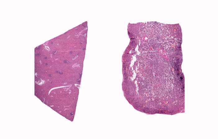
| 15:10, 20 August 2013 | IPLab5Gaucher3.jpg (file) |  |
24 KB | Peter Anderson | This is a low-power photomicrograph of normal spleen (left) and the spleen from this case (right). The loose appearance of the tissue in the Gaucher spleen is due to artifactual loss of tissue during histologic processing. | 1 |
| 15:08, 20 August 2013 | IPLab5Gaucher2.jpg (file) |  |
72 KB | Peter Anderson | This is a cut section of spleen from this case. Again note the fine granular appearance to the tissue. | 1 |
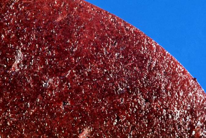
| 15:07, 20 August 2013 | IPLab5Gaucher1.jpg (file) |  |
60 KB | Peter Anderson | This is a gross photograph of spleen from this case. The spleen is enlarged and the surface is finely granular. | 1 |
| 14:57, 20 August 2013 | IPLab5Hemochromatosis11.jpg (file) |  |
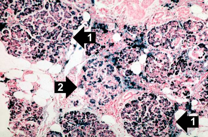
81 KB | Peter Anderson | This is a histologic section of pancreas from this case stained for iron (Prussian blue). Note the accumulation of iron in the parenchymal cells (1). There is also iron in the pancreatic islets (2). | 1 |
| 14:56, 20 August 2013 | IPLab5Hemochromatosis10.jpg (file) |  |
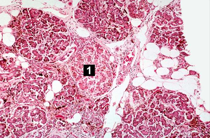
91 KB | Peter Anderson | This is a histologic section of pancreas from this case. It is difficult to appreciate at this magnification, but there is brown pigment in the pancreatic acinar cells. Note the islets of Langerhans (1). | 1 |
| 14:56, 20 August 2013 | IPLab5Hemochromatosis9.jpg (file) |  |
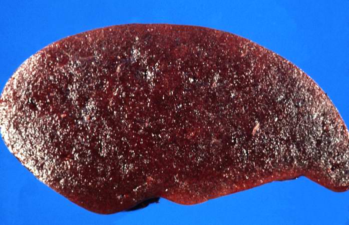
55 KB | Peter Anderson | This is a gross picture of pancreas from this case. Note the brown discoloration of the tissue. | 1 |
| 14:55, 20 August 2013 | IPLab5Hemochromatosis8.jpg (file) |  |
78 KB | Peter Anderson | This higher-power view of liver stained with Prussian blue demonstrates the marked accumulation of iron within the parenchymal cells (1) and in the Kupffer cells in the periportal area (2). | 1 |
| 14:55, 20 August 2013 | IPLab5Hemochromatosis7.jpg (file) |  |
24 KB | Peter Anderson | This is a low-power view of liver section stained with Prussian blue. Prussian blue reacts with iron in the tissue to give a blue color. | 1 |
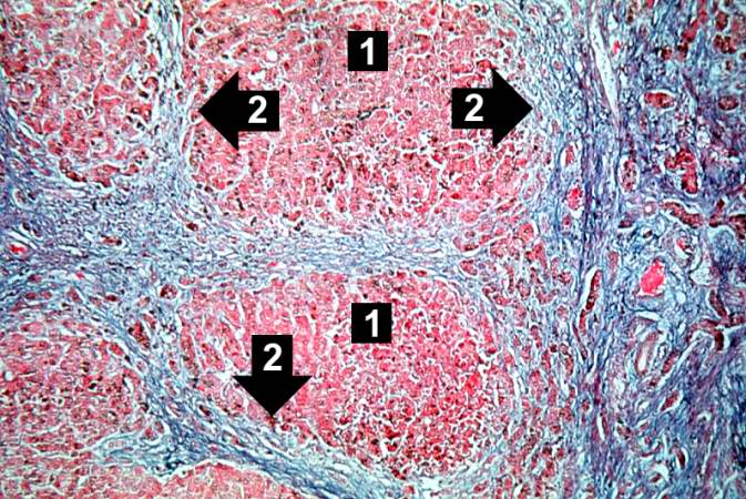
| 14:54, 20 August 2013 | IPLab5Hemochromatosis6.jpg (file) |  |
99 KB | Peter Anderson | This trichrome stain of liver section demonstrates the increased fibrous connective tissue in this liver. Note that the liver nodules (1) are surrounded by fibrous connective tissue (2). | 1 |
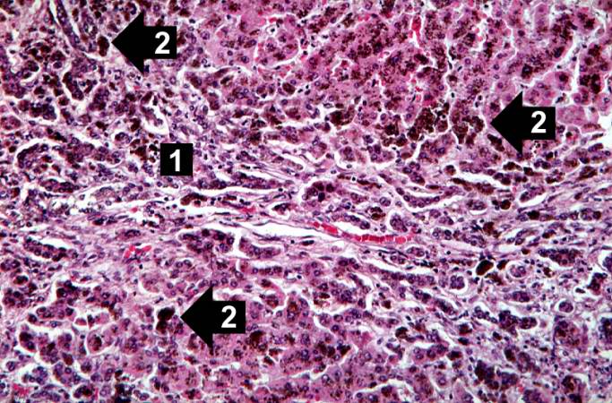
| 14:54, 20 August 2013 | IPLab5Hemochromatosis5.jpg (file) |  |
92 KB | Peter Anderson | This higher-power photomicrograph demonstrates the increased fibrosis in the periportal area (1) and the pigment accumulation (2). | 1 |
| 14:53, 20 August 2013 | IPLab5Hemochromatosis4.jpg (file) |  |
94 KB | Peter Anderson | This higher-power view of liver from this case demonstrates the nodules and the brown/black pigment within liver parenchymal cells (arrows). | 1 |
| 14:53, 20 August 2013 | IPLab5Hemochromatosis3.jpg (file) |  |
23 KB | Peter Anderson | This is a low-power micrograph of liver from this patient. Note the nodularity of the tissue (arrows). | 1 |