File list
This special page shows all uploaded files.
| Date | Name | Thumbnail | Size | User | Description | Versions |
|---|---|---|---|---|---|---|
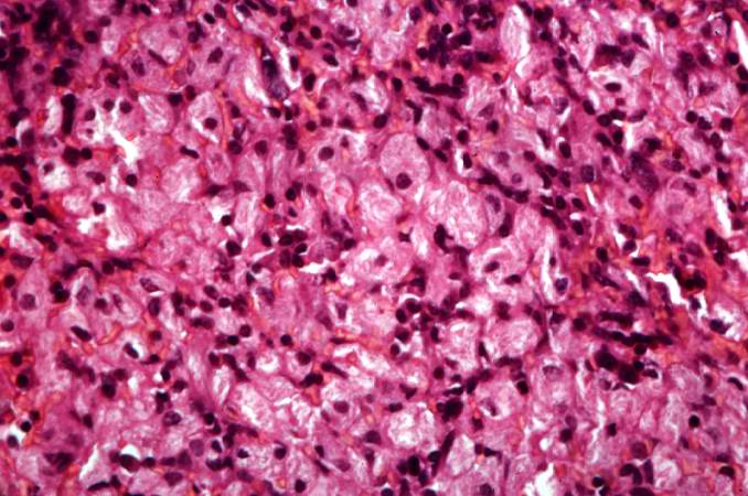
| 15:12, 20 August 2013 | IPLab5Gaucher7.jpg (file) |  |
64 KB | Peter Anderson | This is another high-power photomicrograph of the spleen from this case. At this high power individual cells can be better appreciated. | 1 |
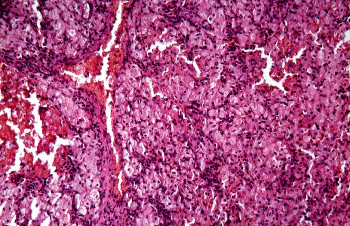
| 15:11, 20 August 2013 | IPLab5Gaucher6.jpg (file) |  |
87 KB | Peter Anderson | This is another high-power photomicrograph of the spleen from this case. At this power it is easier to see the large eosinophilic cells. | 1 |
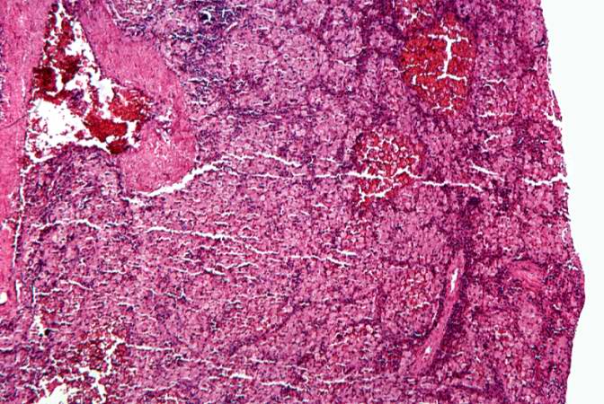
| 15:11, 20 August 2013 | IPLab5Gaucher5.jpg (file) |  |
90 KB | Peter Anderson | This is a higher-power photomicrograph of the spleen from this case. Again there is no white pulp and the red pulp is filled with large eosinophilic cells. | 1 |
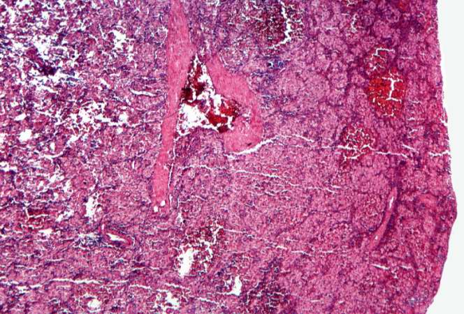
| 15:11, 20 August 2013 | IPLab5Gaucher4.jpg (file) |  |
86 KB | Peter Anderson | This is a photomicrograph of the spleen from this case. There is very little if any white pulp evident in this section. | 1 |
| 15:10, 20 August 2013 | IPLab5Gaucher3.jpg (file) |  |
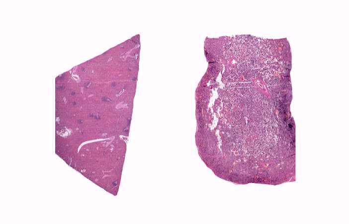
24 KB | Peter Anderson | This is a low-power photomicrograph of normal spleen (left) and the spleen from this case (right). The loose appearance of the tissue in the Gaucher spleen is due to artifactual loss of tissue during histologic processing. | 1 |
| 15:08, 20 August 2013 | IPLab5Gaucher2.jpg (file) |  |
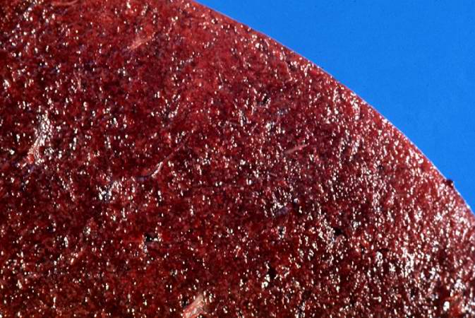
72 KB | Peter Anderson | This is a cut section of spleen from this case. Again note the fine granular appearance to the tissue. | 1 |
| 15:07, 20 August 2013 | IPLab5Gaucher1.jpg (file) |  |
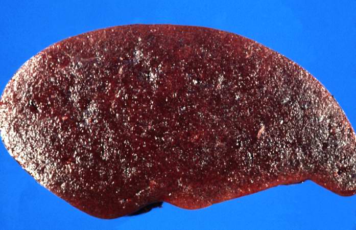
60 KB | Peter Anderson | This is a gross photograph of spleen from this case. The spleen is enlarged and the surface is finely granular. | 1 |
| 14:57, 20 August 2013 | IPLab5Hemochromatosis11.jpg (file) |  |
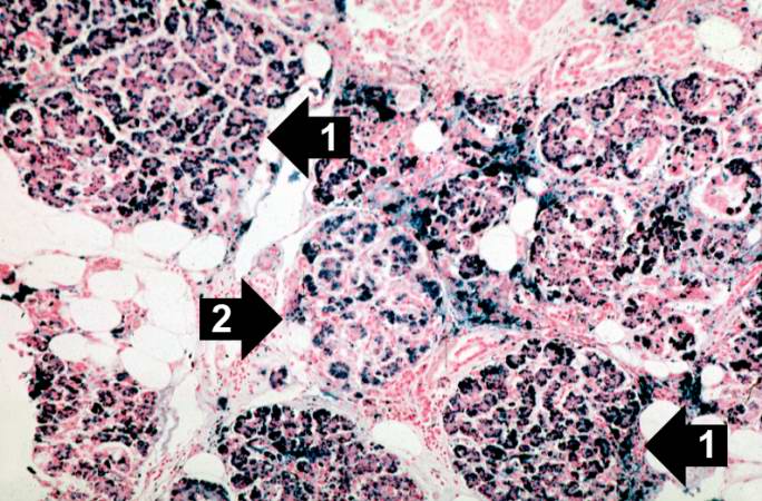
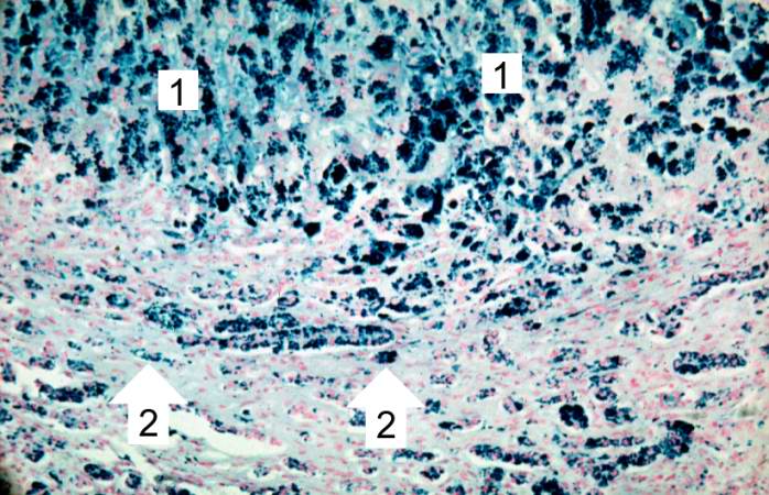
81 KB | Peter Anderson | This is a histologic section of pancreas from this case stained for iron (Prussian blue). Note the accumulation of iron in the parenchymal cells (1). There is also iron in the pancreatic islets (2). | 1 |
| 14:56, 20 August 2013 | IPLab5Hemochromatosis10.jpg (file) |  |
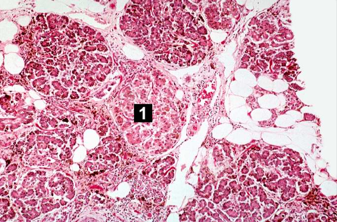
91 KB | Peter Anderson | This is a histologic section of pancreas from this case. It is difficult to appreciate at this magnification, but there is brown pigment in the pancreatic acinar cells. Note the islets of Langerhans (1). | 1 |
| 14:56, 20 August 2013 | IPLab5Hemochromatosis9.jpg (file) |  |
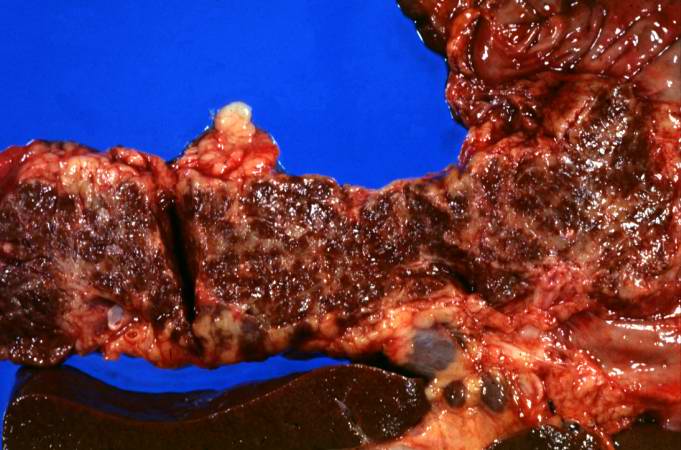
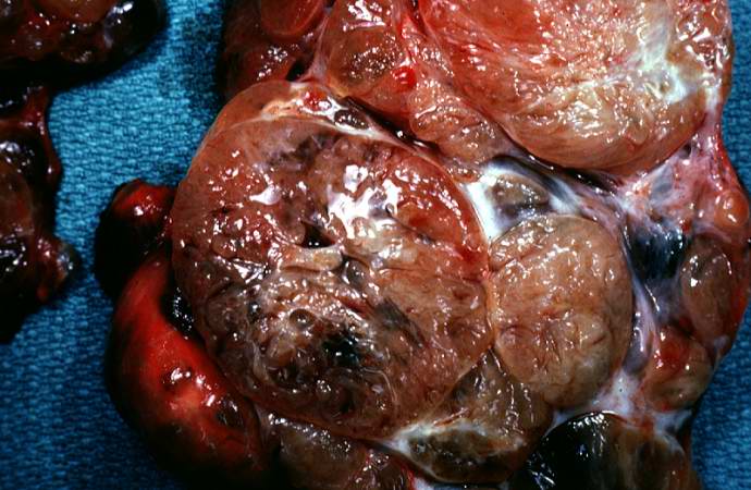
55 KB | Peter Anderson | This is a gross picture of pancreas from this case. Note the brown discoloration of the tissue. | 1 |
| 14:55, 20 August 2013 | IPLab5Hemochromatosis8.jpg (file) |  |
78 KB | Peter Anderson | This higher-power view of liver stained with Prussian blue demonstrates the marked accumulation of iron within the parenchymal cells (1) and in the Kupffer cells in the periportal area (2). | 1 |
| 14:55, 20 August 2013 | IPLab5Hemochromatosis7.jpg (file) |  |
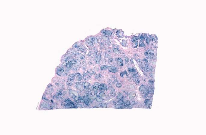
24 KB | Peter Anderson | This is a low-power view of liver section stained with Prussian blue. Prussian blue reacts with iron in the tissue to give a blue color. | 1 |
| 14:54, 20 August 2013 | IPLab5Hemochromatosis6.jpg (file) |  |
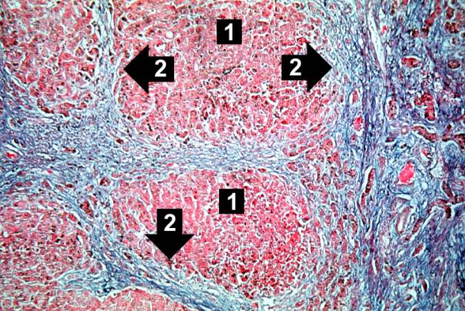
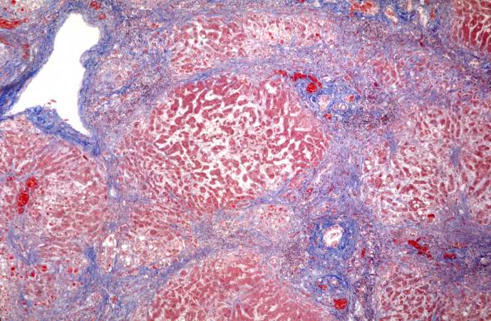
99 KB | Peter Anderson | This trichrome stain of liver section demonstrates the increased fibrous connective tissue in this liver. Note that the liver nodules (1) are surrounded by fibrous connective tissue (2). | 1 |
| 14:54, 20 August 2013 | IPLab5Hemochromatosis5.jpg (file) |  |
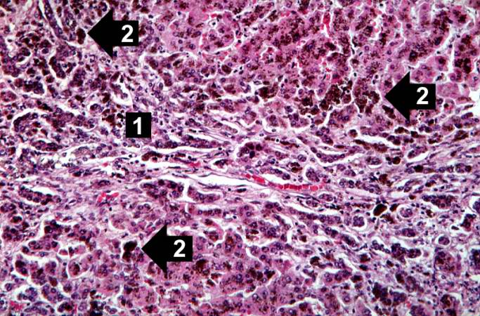
92 KB | Peter Anderson | This higher-power photomicrograph demonstrates the increased fibrosis in the periportal area (1) and the pigment accumulation (2). | 1 |
| 14:53, 20 August 2013 | IPLab5Hemochromatosis4.jpg (file) |  |
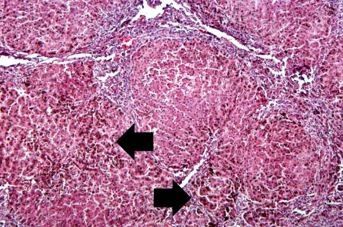
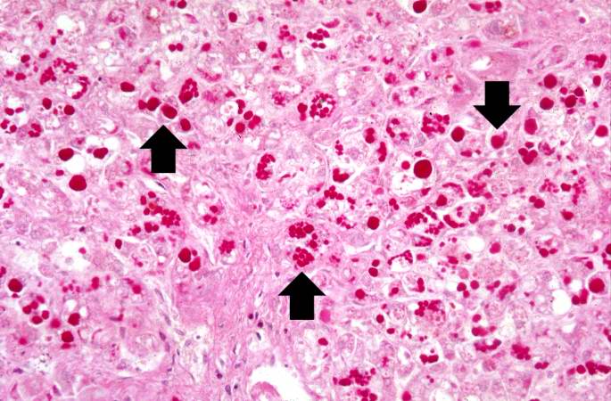
94 KB | Peter Anderson | This higher-power view of liver from this case demonstrates the nodules and the brown/black pigment within liver parenchymal cells (arrows). | 1 |
| 14:53, 20 August 2013 | IPLab5Hemochromatosis3.jpg (file) |  |
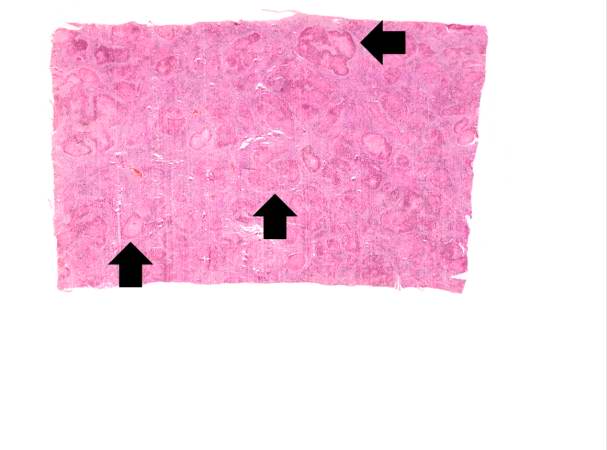
23 KB | Peter Anderson | This is a low-power micrograph of liver from this patient. Note the nodularity of the tissue (arrows). | 1 |
| 14:53, 20 August 2013 | IPLab5Hemochromatosis2.jpg (file) |  |
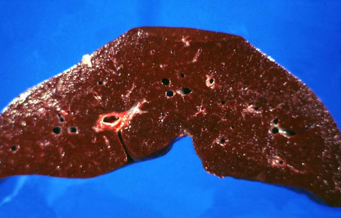
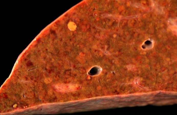
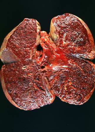
37 KB | Peter Anderson | This is a gross photograph of a cut section of liver from this case of hemochromatosis. Note that the liver is dark brown. Although hard to appreciate in a photograph, the tissue is also firm (cirrhotic). | 1 |
| 14:52, 20 August 2013 | IPLab5Hemochromatosis1.jpg (file) |  |
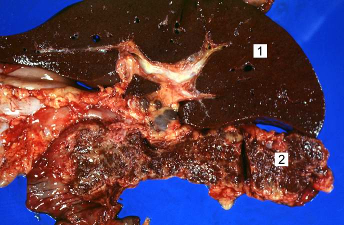
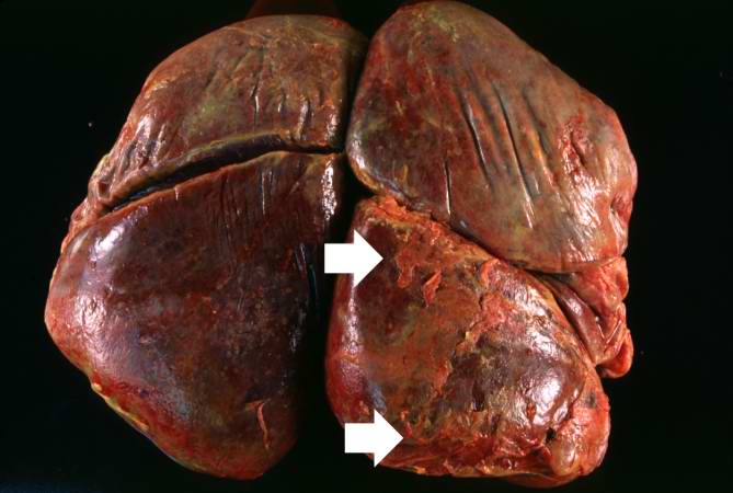
59 KB | Peter Anderson | This is a gross photograph of liver (1) and pancreas (2) from this case of hemochromatosis. Note that both of these organs have a dark brown coloration. | 1 |
| 18:29, 19 August 2013 | IPLab5Downs6.jpg (file) |  |
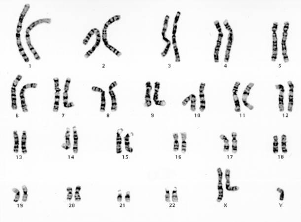
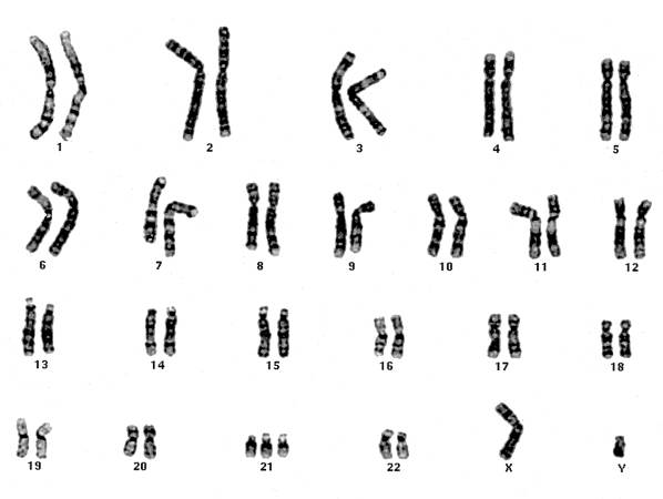
20 KB | Seung Park | This is a karyotype of a patient with Turner syndrome (45, X). | 1 |
| 18:28, 19 August 2013 | IPLab5Downs5.jpg (file) |  |
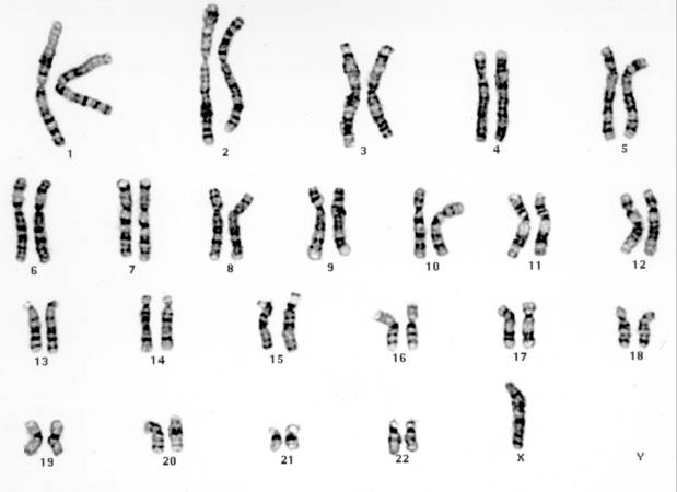
19 KB | Seung Park | This is a karyotype of a patient with Klinefelter syndrome (47, XXY). | 1 |
| 18:28, 19 August 2013 | IPLab5Antitrypsin12.jpg (file) |  |
68 KB | Peter Anderson | This is a high-power photomicrograph of liver stained with periodic-acid Schiff's (PAS) stain. This demonstrates the PAS-positive granules of defective alpha 1-antitrypsin that accumulate in the Golgi of hepatocytes (arrows). | 1 |
| 18:28, 19 August 2013 | IPLab5Downs4.jpg (file) |  |
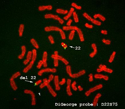
22 KB | Seung Park | FISH is also useful in the diagnosis of other genetic disorders. This is an example of FISH staining on another patient using a probe specific for DiGeorge's disease. The arrow shows that there is a deletion on chromosome 22, which is diagnostic for Di... | 1 |
| 18:28, 19 August 2013 | IPLab5Antitrypsin11.jpg (file) |  |
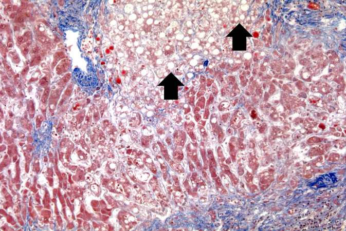
77 KB | Peter Anderson | This is a higher-power photomicrograph of a trichrome-stained section of liver. This section demonstrates the fibrosis (blue material) and the fatty change (arrows). | 1 |
| 18:28, 19 August 2013 | IPLab5Downs3.jpg (file) |  |
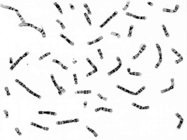
21 KB | Seung Park | Chromosomes from the chromosome spread are lined up to demonstrate the karyotype. In this case there are three copies of chromosome 21, just as noted in the FISH. | 1 |
| 18:27, 19 August 2013 | IPLab5Antitrypsin10.jpg (file) |  |
80 KB | Peter Anderson | This is a low-power photomicrograph of a trichrome-stained section of liver. There is bridging fibrosis (blue material) between portal regions. | 1 |
| 18:27, 19 August 2013 | IPLab5Downs2.jpg (file) |  |
17 KB | Seung Park | These cells, obtained by amniocentesis, were cultured and then arrested in metaphase. Nuclei from these cells were isolated and stained to demonstrate the banding pattern of each chromosome. This photograph shows a "chromosome spread." Each chromosome ... | 1 |
| 18:27, 19 August 2013 | IPLab5Downs1.jpg (file) |  |
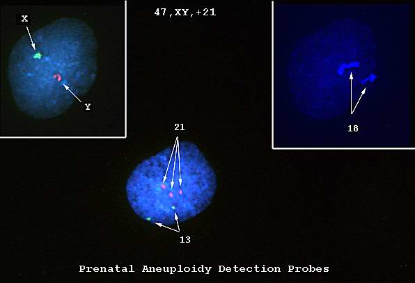
18 KB | Seung Park | This is a photomicrograph of cells obtained by amniocentesis that were stained using FISH. The cell in panel 1 was stained with markers specific for the X and Y-chromosomes. The cell in panel 2 was stained with a marker specific for chromosome 18. The ... | 1 |
| 18:27, 19 August 2013 | IPLab5Antitrypsin9.jpg (file) |  |
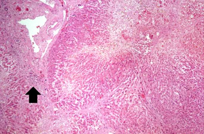
70 KB | Peter Anderson | This is a low-power photomicrograph of an H&E-stained section of liver. There are increased numbers of inflammatory cells in the periportal region (arrow) and the central vein areas are pale. | 1 |
| 18:26, 19 August 2013 | IPLab5Antitrypsin8.jpg (file) |  |
35 KB | Peter Anderson | This is a closer view of the cut section of liver from this case. There is a definite micronodular pattern to the liver parenchyma. | 1 |
| 18:26, 19 August 2013 | IPLab5Antitrypsin7.jpg (file) |  |
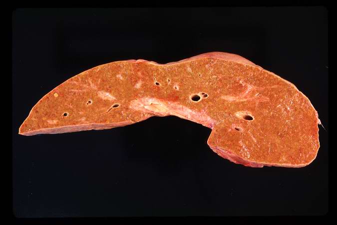
25 KB | Peter Anderson | This is a gross photograph of the cut section of liver from this case. In this view the liver looks smaller than normal and there is a definite micronodular appearance. | 1 |
| 18:25, 19 August 2013 | IPLab5Antitrypsin6.jpg (file) |  |
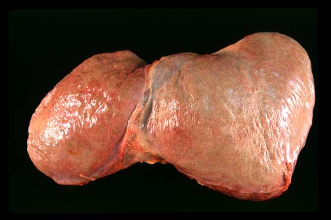
29 KB | Peter Anderson | This is a gross photograph of the liver from this case. The capsule is somewhat thickened and the surface is slightly roughened, though it is difficult to appreciate the nodularity of the liver. | 1 |
| 18:23, 19 August 2013 | IPLab6GravesDisease7.jpg (file) |  |
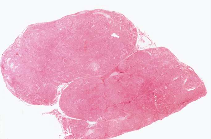
61 KB | Seung Park | Closer view of cut surface of the thyroid with nodular goiter. Note the multilobular appearance of the tissue. | 1 |
| 18:23, 19 August 2013 | IPLab6GravesDisease6.jpg (file) |  |
63 KB | Seung Park | This is a gross photograph of a thyroid from a case of nodular goiter. | 1 |
| 18:23, 19 August 2013 | IPLab6GravesDisease5.jpg (file) |  |
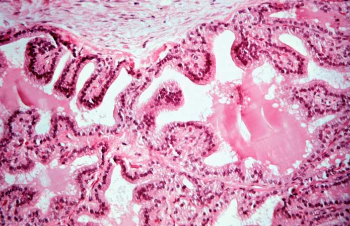
65 KB | Seung Park | This is a high-power photomicrograph of thyroid. Note the papillary projections and the moth-eaten appearance of the colloid. This appearance indicates active absorption of the colloid to form thyroglobulin. | 1 |
| 18:22, 19 August 2013 | IPLab6GravesDisease4.jpg (file) |  |
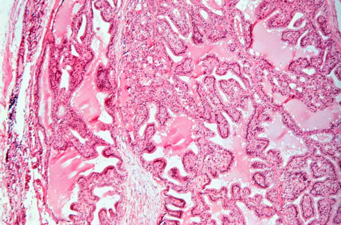
77 KB | Seung Park | This is a high-power photomicrograph of thyroid. Note the cellularity of the tissue with marked infolding of the epithelial tissue. | 1 |
| 18:22, 19 August 2013 | IPLab6GravesDisease3.jpg (file) |  |
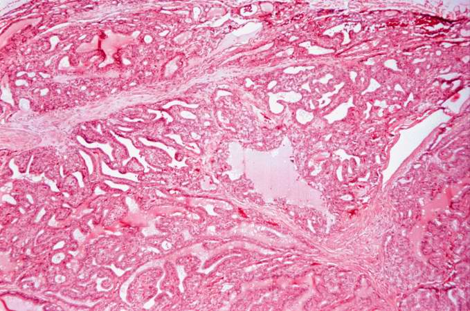
75 KB | Seung Park | This is a higher-power photomicrograph of thyroid. The tissue is very cellular and there is little colloid. | 1 |
| 18:22, 19 August 2013 | IPLab6GravesDisease2.jpg (file) |  |
29 KB | Seung Park | This is a low-power photomicrograph of thyroid tissue from this case. The tissue is very cellular with very little colloid. | 1 |
| 18:21, 19 August 2013 | IPLab6GravesDisease1.jpg (file) |  |
43 KB | Seung Park | This is a photograph of the thyroid from this case. Note that the gland is enlarged and is dark red. | 1 |
| 18:13, 19 August 2013 | IPLab5Antitrypsin3.jpg (file) |  |
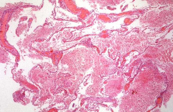
67 KB | Peter Anderson | This is a gross photograph of the bronchi and lungs. Note the hemorrhage in the bronchi and in the lung parenchyma. | 1 |
| 18:09, 19 August 2013 | IPLab6RA10.jpg (file) |  |
65 KB | Seung Park | This is a high-power photomicrograph of another region with macrophages (right), fibrocytes (left), and occasional lymphocytes throughout the lesion. | 1 |
| 18:09, 19 August 2013 | IPLab5Antitrypsin5.jpg (file) |  |
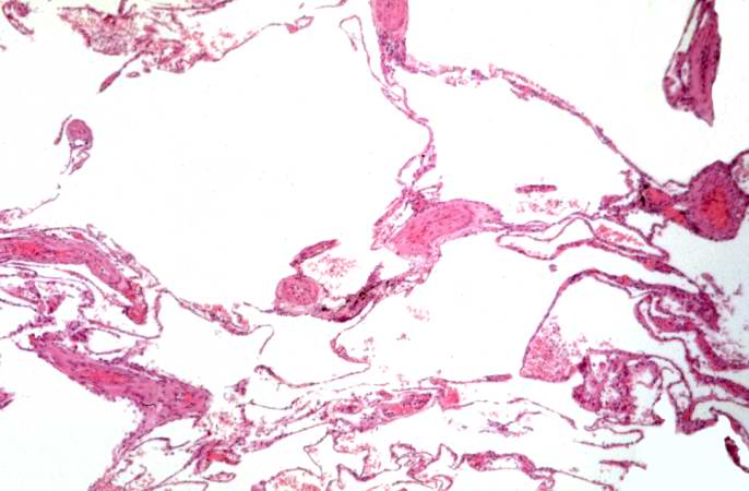
41 KB | Peter Anderson | This is a low-power photomicrograph from an area of the lung without significant hemorrhage. The enlarged, emphysematous air spaces are easily appreciated. | 1 |
| 18:08, 19 August 2013 | IPLab6RA9.jpg (file) |  |
63 KB | Seung Park | This is a high-power photomicrograph of the mononuclear cells which surround the central area of necrosis. The focal accumulations of fibrinoid material are clearly visible. Lymphocytes are present in the extreme right of this image. | 1 |
| 18:08, 19 August 2013 | IPLab6RA8.jpg (file) |  |
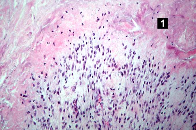
59 KB | Seung Park | This higher-power photomicrograph of the tissue illustrates the palisading nuclei of the monocytes which are located around the periphery of the central necrotic region (1). | 1 |
| 18:07, 19 August 2013 | IPLab6RA7.jpg (file) |  |
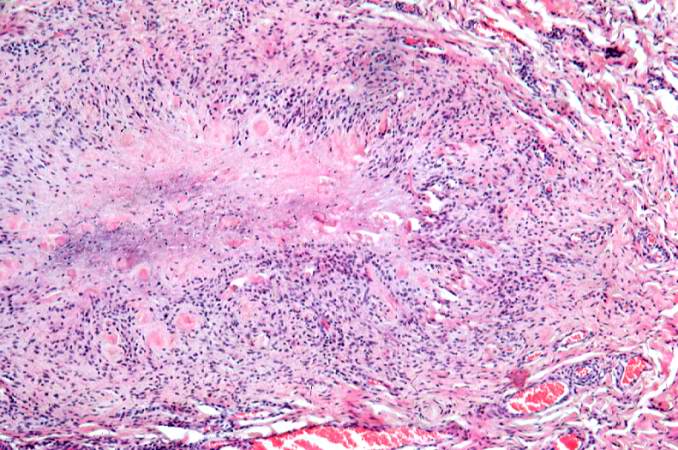
82 KB | Seung Park | This higher-power photomicrograph of the subcutaneous nodule again demonstrates the necrotic center and peripheral rim of macrophages, fibrocytes, and occasional lymphocytes. There are focal accumulations of hyaline material (fibrinoid material) within... | 1 |
| 18:07, 19 August 2013 | IPLab5Antitrypsin4.jpg (file) |  |
69 KB | Peter Anderson | This is a gross photograph of the bronchi and lungs. Note the hemorrhage in the bronchi and in the lung parenchyma. | 1 |
| 18:07, 19 August 2013 | IPLab6RA6.jpg (file) |  |
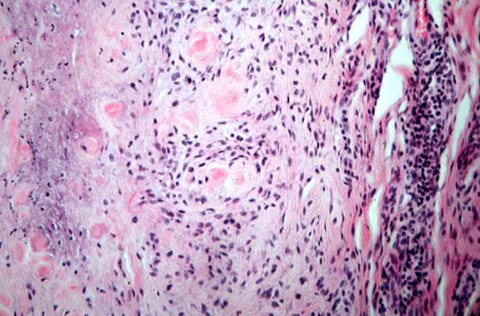
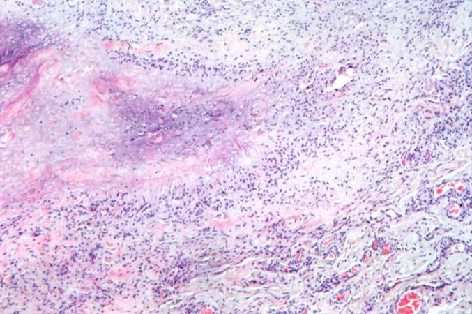
64 KB | Seung Park | This higher-power photomicrograph of the subcutaneous nodule shows a granulomatous lesion with a necrotic center and a peripheral rim of macrophages, fibrocytes, and occasional lymphocytes. In the necrotic center of the granuloma there is some minerali... | 1 |
| 18:07, 19 August 2013 | IPLab5Antitrypsin2.jpg (file) |  |
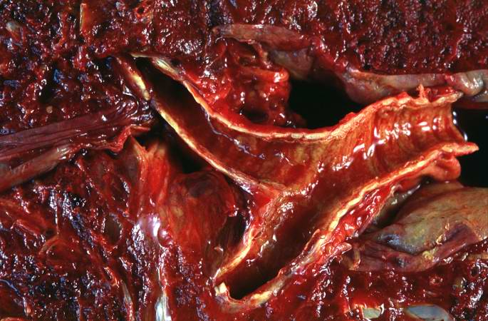
32 KB | Peter Anderson | This is a gross photograph of the cut sections of lung from this case. The lung parenchyma is markedly hemorrhagic and consolidated. Again the hemorrhage makes it difficult to appreciate the emphysematous changes. | 1 |
| 18:07, 19 August 2013 | IPLab6RA5.jpg (file) |  |
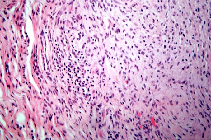
11 KB | Seung Park | This is a low-power photomicrograph of the subcutaneous nodule from this patient. | 1 |
| 18:06, 19 August 2013 | IPLab5Antitrypsin1.jpg (file) |  |
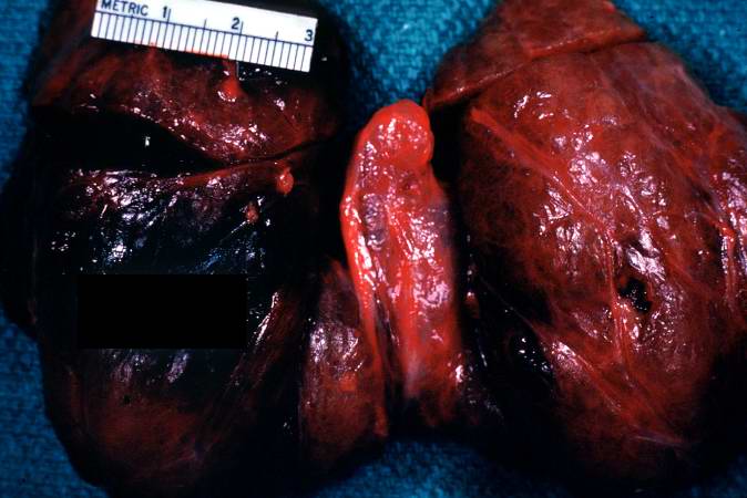
37 KB | Peter Anderson | This is a gross photograph of the lungs from this case. The rough friable material on the surface of the lung (arrows) is fibrinous exudate and fibrous tissue. This reaction on the surface of the lung is due to the recent surgery. The emphysematous cha... | 1 |
| 18:06, 19 August 2013 | IPLab6RA4.jpg (file) |  |
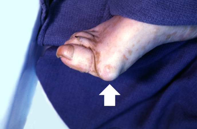
24 KB | Seung Park | This is a gross photograph of the foot from this same patient. Note the subcutaneous nodule on the medial aspect of the foot (arrow). | 1 |