File list
This special page shows all uploaded files.
| Date | Name | Thumbnail | Size | Description | Versions |
|---|---|---|---|---|---|
| 14:56, 20 August 2013 | IPLab5Hemochromatosis10.jpg (file) |  |
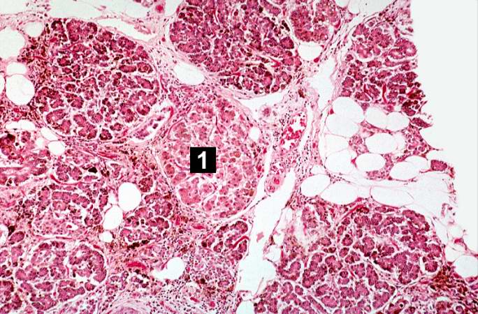
91 KB | This is a histologic section of pancreas from this case. It is difficult to appreciate at this magnification, but there is brown pigment in the pancreatic acinar cells. Note the islets of Langerhans (1). | 1 |
| 14:56, 20 August 2013 | IPLab5Hemochromatosis9.jpg (file) |  |
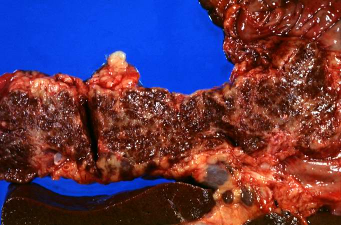
55 KB | This is a gross picture of pancreas from this case. Note the brown discoloration of the tissue. | 1 |
| 14:55, 20 August 2013 | IPLab5Hemochromatosis8.jpg (file) |  |
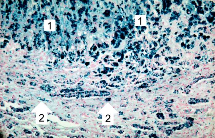
78 KB | This higher-power view of liver stained with Prussian blue demonstrates the marked accumulation of iron within the parenchymal cells (1) and in the Kupffer cells in the periportal area (2). | 1 |
| 14:55, 20 August 2013 | IPLab5Hemochromatosis7.jpg (file) |  |
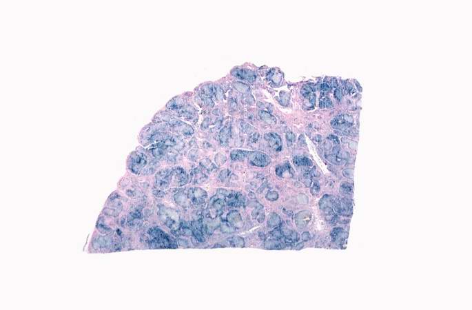
24 KB | This is a low-power view of liver section stained with Prussian blue. Prussian blue reacts with iron in the tissue to give a blue color. | 1 |
| 14:54, 20 August 2013 | IPLab5Hemochromatosis6.jpg (file) |  |
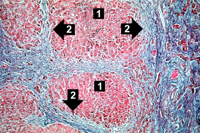
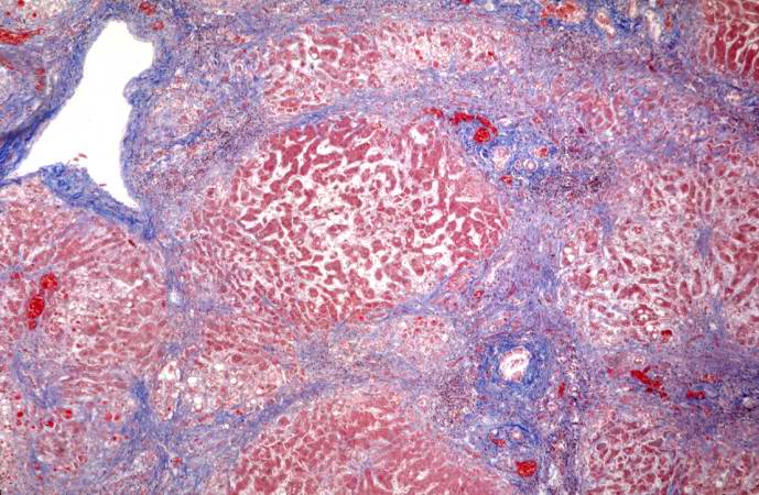
99 KB | This trichrome stain of liver section demonstrates the increased fibrous connective tissue in this liver. Note that the liver nodules (1) are surrounded by fibrous connective tissue (2). | 1 |
| 14:54, 20 August 2013 | IPLab5Hemochromatosis5.jpg (file) |  |
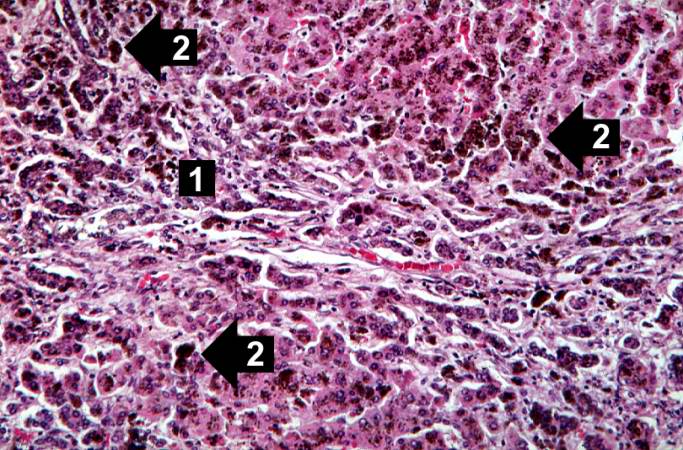
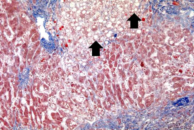
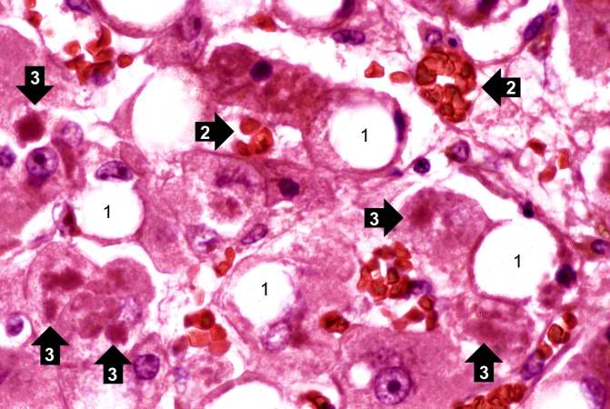
92 KB | This higher-power photomicrograph demonstrates the increased fibrosis in the periportal area (1) and the pigment accumulation (2). | 1 |
| 14:53, 20 August 2013 | IPLab5Hemochromatosis4.jpg (file) |  |
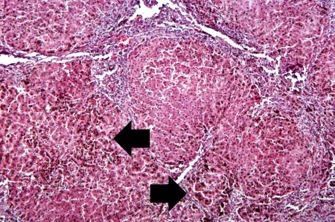
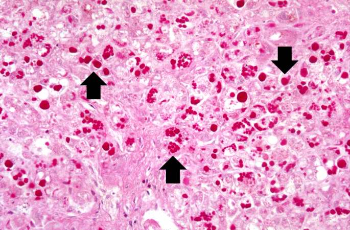
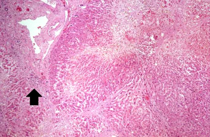
94 KB | This higher-power view of liver from this case demonstrates the nodules and the brown/black pigment within liver parenchymal cells (arrows). | 1 |
| 14:53, 20 August 2013 | IPLab5Hemochromatosis3.jpg (file) |  |
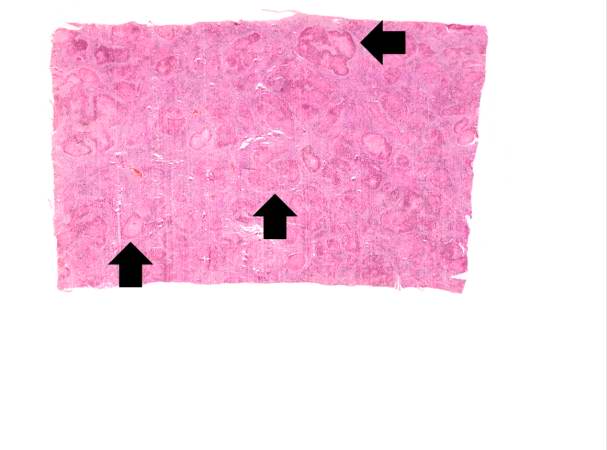
23 KB | This is a low-power micrograph of liver from this patient. Note the nodularity of the tissue (arrows). | 1 |
| 14:53, 20 August 2013 | IPLab5Hemochromatosis2.jpg (file) |  |
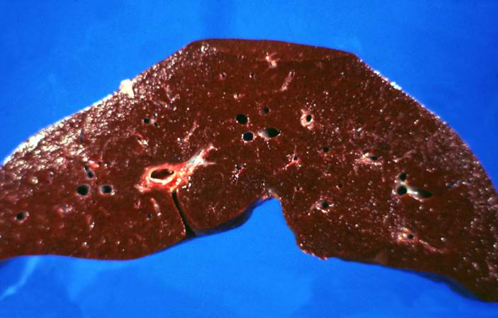
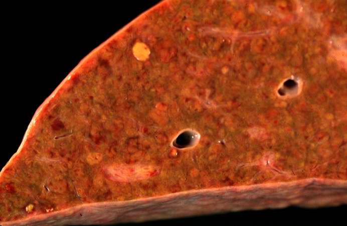
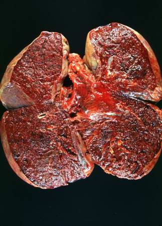
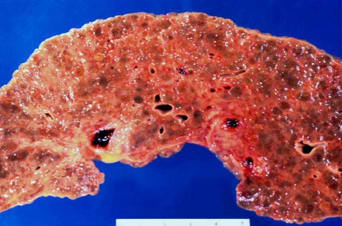
37 KB | This is a gross photograph of a cut section of liver from this case of hemochromatosis. Note that the liver is dark brown. Although hard to appreciate in a photograph, the tissue is also firm (cirrhotic). | 1 |
| 14:52, 20 August 2013 | IPLab5Hemochromatosis1.jpg (file) |  |
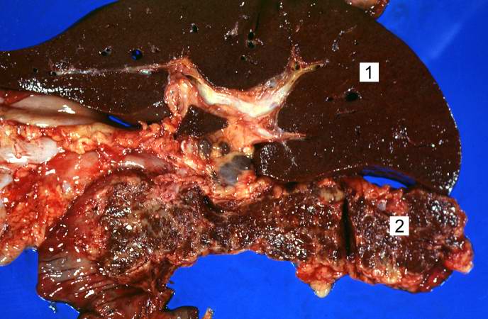
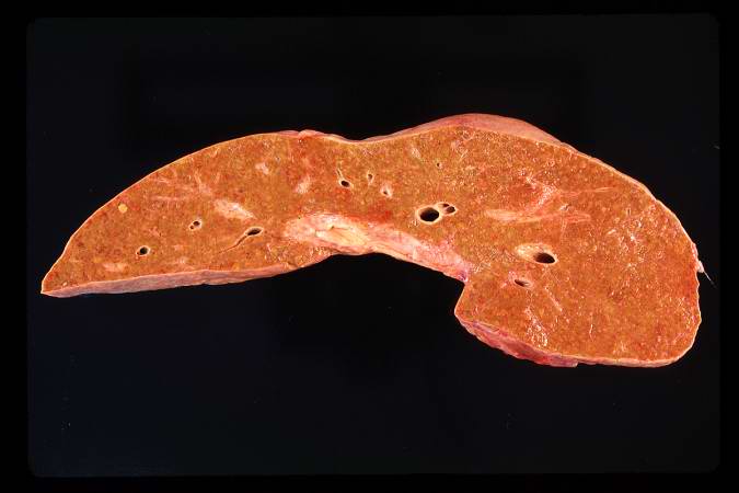
59 KB | This is a gross photograph of liver (1) and pancreas (2) from this case of hemochromatosis. Note that both of these organs have a dark brown coloration. | 1 |
| 18:28, 19 August 2013 | IPLab5Antitrypsin12.jpg (file) |  |
68 KB | This is a high-power photomicrograph of liver stained with periodic-acid Schiff's (PAS) stain. This demonstrates the PAS-positive granules of defective alpha 1-antitrypsin that accumulate in the Golgi of hepatocytes (arrows). | 1 |
| 18:28, 19 August 2013 | IPLab5Antitrypsin11.jpg (file) |  |
77 KB | This is a higher-power photomicrograph of a trichrome-stained section of liver. This section demonstrates the fibrosis (blue material) and the fatty change (arrows). | 1 |
| 18:27, 19 August 2013 | IPLab5Antitrypsin10.jpg (file) |  |
80 KB | This is a low-power photomicrograph of a trichrome-stained section of liver. There is bridging fibrosis (blue material) between portal regions. | 1 |
| 18:27, 19 August 2013 | IPLab5Antitrypsin9.jpg (file) |  |
70 KB | This is a low-power photomicrograph of an H&E-stained section of liver. There are increased numbers of inflammatory cells in the periportal region (arrow) and the central vein areas are pale. | 1 |
| 18:26, 19 August 2013 | IPLab5Antitrypsin8.jpg (file) |  |
35 KB | This is a closer view of the cut section of liver from this case. There is a definite micronodular pattern to the liver parenchyma. | 1 |
| 18:26, 19 August 2013 | IPLab5Antitrypsin7.jpg (file) |  |
25 KB | This is a gross photograph of the cut section of liver from this case. In this view the liver looks smaller than normal and there is a definite micronodular appearance. | 1 |
| 18:25, 19 August 2013 | IPLab5Antitrypsin6.jpg (file) |  |
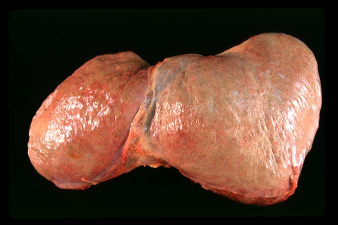
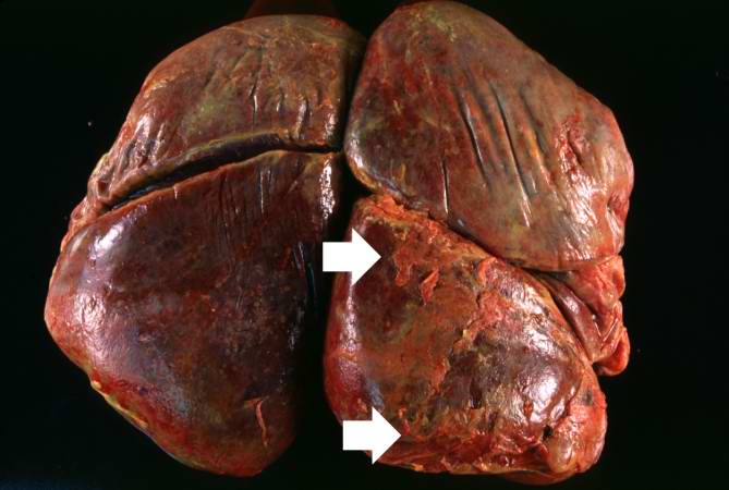
29 KB | This is a gross photograph of the liver from this case. The capsule is somewhat thickened and the surface is slightly roughened, though it is difficult to appreciate the nodularity of the liver. | 1 |
| 18:13, 19 August 2013 | IPLab5Antitrypsin3.jpg (file) |  |
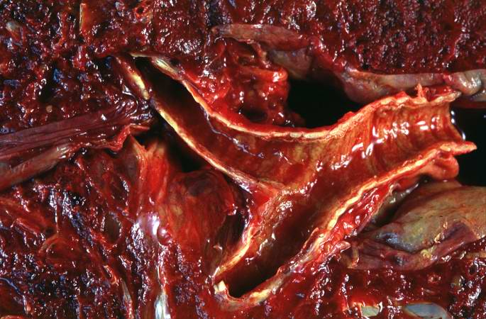
67 KB | This is a gross photograph of the bronchi and lungs. Note the hemorrhage in the bronchi and in the lung parenchyma. | 1 |
| 18:09, 19 August 2013 | IPLab5Antitrypsin5.jpg (file) |  |
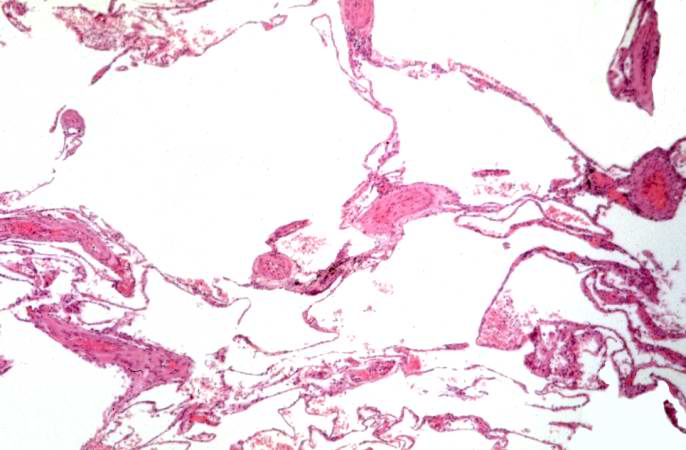
41 KB | This is a low-power photomicrograph from an area of the lung without significant hemorrhage. The enlarged, emphysematous air spaces are easily appreciated. | 1 |
| 18:07, 19 August 2013 | IPLab5Antitrypsin4.jpg (file) |  |
69 KB | This is a gross photograph of the bronchi and lungs. Note the hemorrhage in the bronchi and in the lung parenchyma. | 1 |
| 18:07, 19 August 2013 | IPLab5Antitrypsin2.jpg (file) |  |
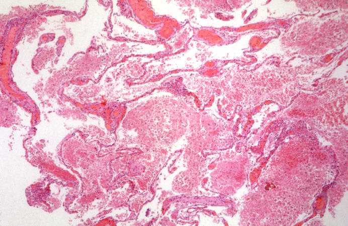
32 KB | This is a gross photograph of the cut sections of lung from this case. The lung parenchyma is markedly hemorrhagic and consolidated. Again the hemorrhage makes it difficult to appreciate the emphysematous changes. | 1 |
| 18:06, 19 August 2013 | IPLab5Antitrypsin1.jpg (file) |  |
37 KB | This is a gross photograph of the lungs from this case. The rough friable material on the surface of the lung (arrows) is fibrinous exudate and fibrous tissue. This reaction on the surface of the lung is due to the recent surgery. The emphysematous cha... | 1 |
| 17:54, 19 August 2013 | IPLab5PolycysticKidney11.jpg (file) |  |
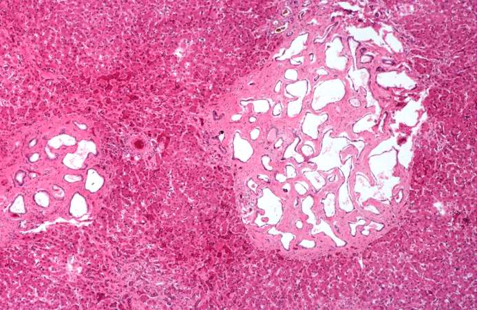
68 KB | This is a higher-power photomicrograph of liver cyst. These cystic structures are lined by biliary epithelium. | 1 |
| 17:54, 19 August 2013 | IPLab5PolycysticKidney10.jpg (file) |  |
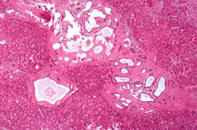
61 KB | This is another photomicrograph of liver demonstrating the histologic appearance of these cysts. These cystic structures are associated with the biliary tree. | 1 |
| 17:53, 19 August 2013 | IPLab5PolycysticKidney9.jpg (file) |  |
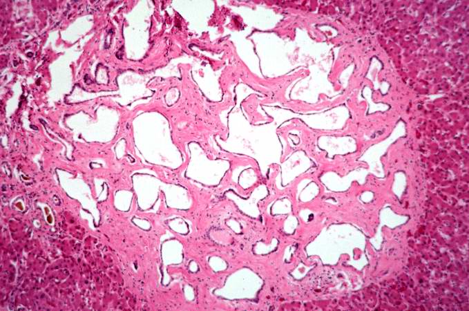
81 KB | This photomicrograph of liver demonstrates the histologic appearance of these cysts. | 1 |
| 17:53, 19 August 2013 | IPLab5PolycysticKidney8.jpg (file) |  |
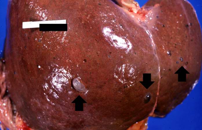
42 KB | This is a gross photograph of the liver from this patient. Multiple cysts can be seen on the surface of this liver (arrows). | 1 |
| 17:52, 19 August 2013 | IPLab5PolycysticKidney7.jpg (file) |  |
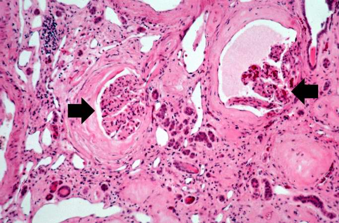
66 KB | This high-power photomicrograph shows abnormal glomeruli (arrows) and some tubules. | 1 |
| 17:52, 19 August 2013 | IPLab5PolycysticKidney6.jpg (file) |  |
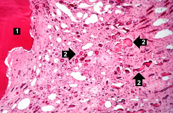
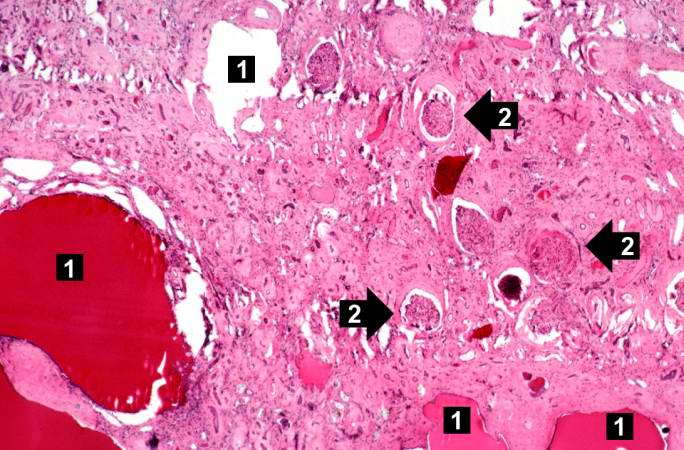
66 KB | This is a higher-power photomicrograph of polycystic kidney showing the edge of a large cyst (1). In this section there are numerous tubules and dilated collecting ducts (2) that are filled with the same red proteinaceous material as the larger cysts. | 1 |
| 17:51, 19 August 2013 | IPLab5PolycysticKidney5.jpg (file) |  |
53 KB | This is another low-power photomicrograph of an H&E-stained section from this polycystic kidney. Again note the large cystic structures (arrows)and the fibrous connective tissue throughout this section. | 1 |
| 17:51, 19 August 2013 | IPLab5PolycysticKidney4.jpg (file) |  |
69 KB | This is a low-power photomicrograph of an H&E-stained section from this polycystic kidney. Note the large cystic structures (1), the few residual glomeruli (2), and the fibrous connective tissue throughout this section. | 1 |
| 17:49, 19 August 2013 | IPLab5PolycysticKidney3.jpg (file) |  |
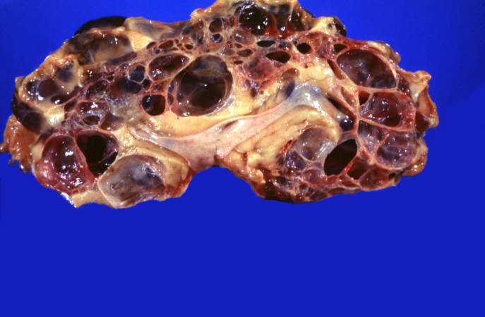
33 KB | This is a gross photograph of a cut section from one of these polycystic kidneys. Note that the renal parenchyma is almost completely replaced by cystic structures. | 1 |
| 17:40, 19 August 2013 | IPLab5PolycysticKidney2.jpg (file) |  |
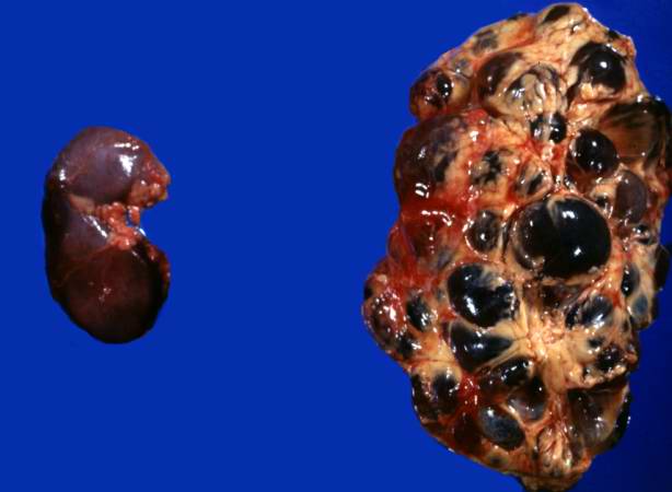
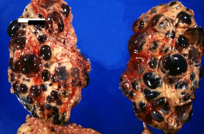
26 KB | This is a gross photograph of the kidneys from this case. Note that both kidneys contain multiple large cysts (arrows). | 1 |
| 17:39, 19 August 2013 | IPLab5PolycysticKidney1.jpg (file) |  |
48 KB | This is a gross photograph of the kidneys from this case. Note that both kidneys contain multiple large cysts (arrows). | 1 |
| 17:22, 19 August 2013 | IPLab5Neurofibromatosis8.jpg (file) |  |
59 KB | This is a high-power photomicrograph of the cells in the neurofibroma. | 1 |
| 17:21, 19 August 2013 | IPLab5Neurofibromatosis7.jpg (file) |  |
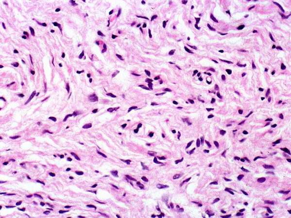
74 KB | This higher-power photomicrograph of the neurofibroma shows more clearly the elongated cells (primarily Schwann cells) that make up this tumor. | 1 |
| 17:21, 19 August 2013 | IPLab5Neurofibromatosis6.jpg (file) |  |
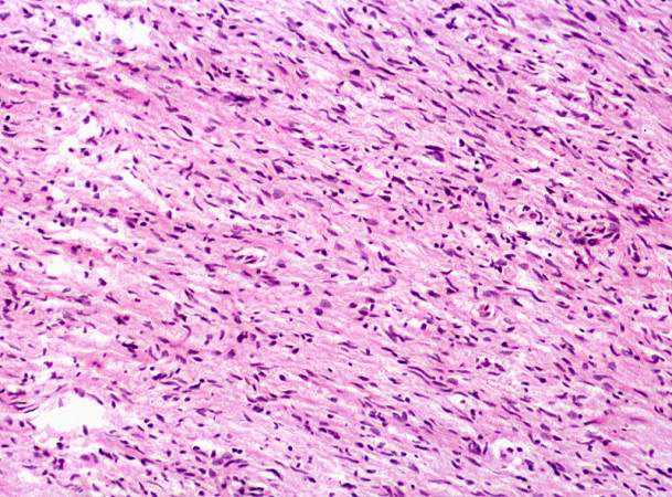
89 KB | This is a higher-power photomicrograph of the neurofibroma demonstrating the loose pattern of elongated cells making up the tumor mass. | 1 |
| 17:20, 19 August 2013 | IPLab5Neurofibromatosis5.jpg (file) |  |
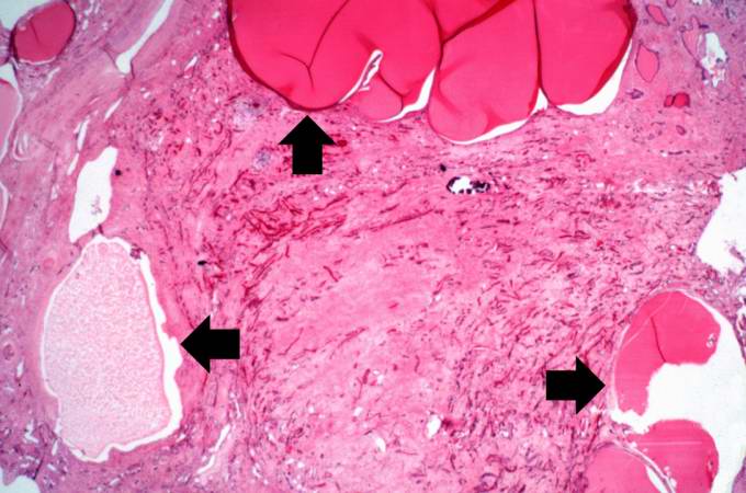
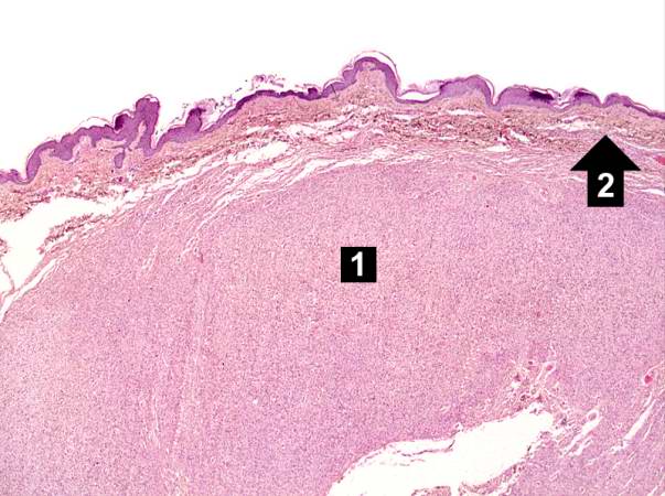
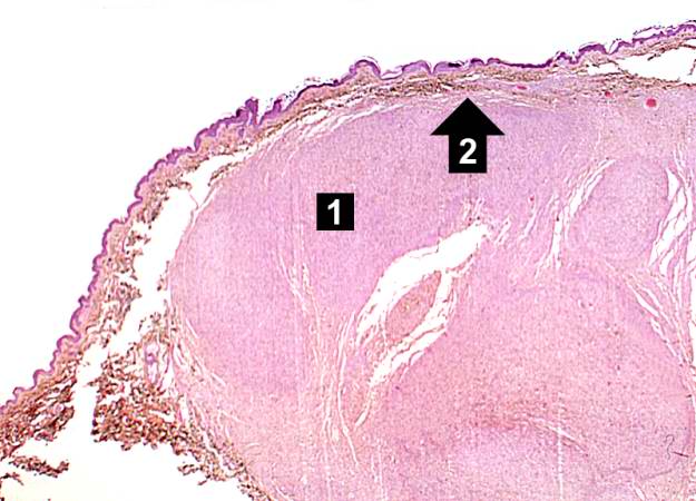
53 KB | This is a higher-power photomicrograph of the neurofibroma (1) with the overlying skin (2). | 1 |
| 17:19, 19 August 2013 | IPLab5Neurofibromatosis4.jpg (file) |  |
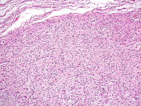
48 KB | This is a low-power photomicrograph of a subcutaneous neurofibroma (1). Note the increased pigmentation in the skin (2). | 1 |
| 17:19, 19 August 2013 | IPLab5Neurofibromatosis3.jpg (file) |  |
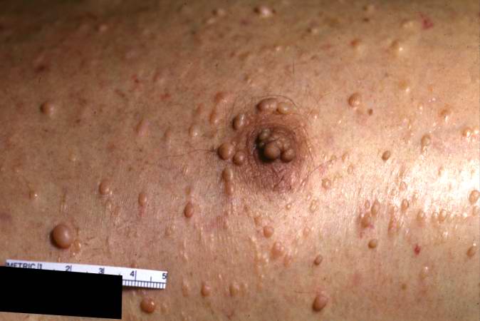
32 KB | This is a closer view of neurofibromas on the skin. | 1 |
| 17:19, 19 August 2013 | IPLab5Neurofibromatosis2.jpg (file) |  |
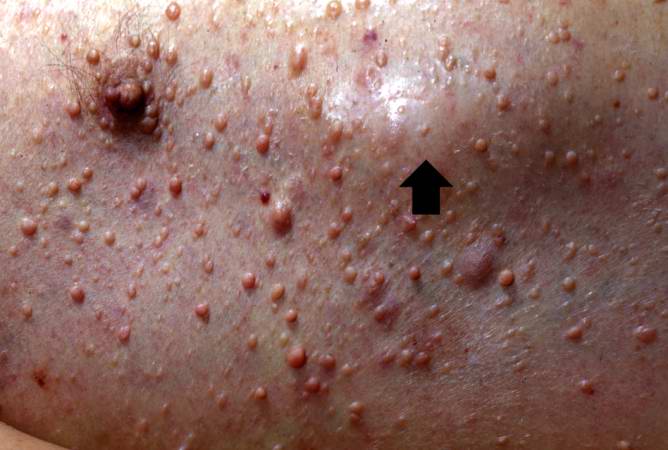
40 KB | This is another view taken at autopsy demonstrating the neurofibromas. Some lesions can be seen as subcutaneous swellings (arrow) and others form pedunculated masses. Most are hyperpigmented. | 1 |
| 17:18, 19 August 2013 | IPLab5Neurofibromatosis1.jpg (file) |  |
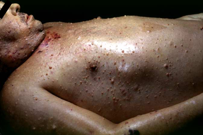
137 KB | This photograph, taken at autopsy, demonstrates the distribution of neurofibromas on the skin of this patient. | 1 |
| 17:02, 19 August 2013 | IPLab2FattyChange12.jpg (file) |  |
49 KB | This is a cut surface of the same tissue seen in the previous slide. Note the marked nodular pattern. The paler-staining areas between the round nodules represent fibrous connective tissue. | 1 |
| 17:00, 19 August 2013 | IPLab2FattyChange11.jpg (file) |  |
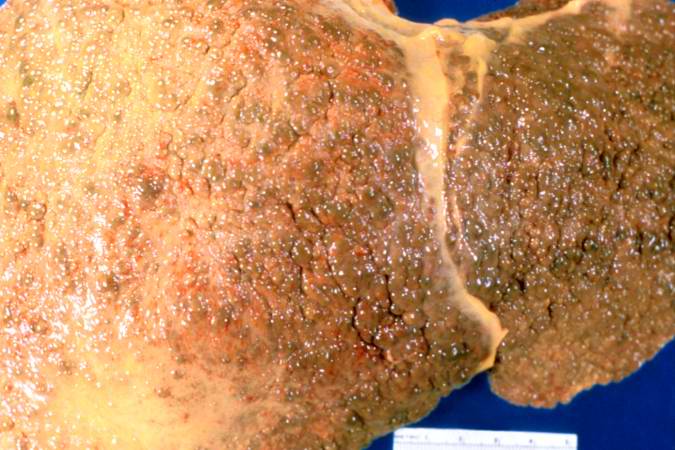
58 KB | This gross photograph of liver demonstrates severe nodular cirrhosis. Note the extensive scarring of the capsule and the nodular projections of tissue through the uncut capsule in this tissue. The green color is due to the accumulation of bile pigment. | 1 |
| 16:59, 19 August 2013 | IPLab2FattyChange9.jpg (file) |  |
58 KB | This photomicrograph of the liver is from another patient with a history of alcohol use. There are some clear vacuoles indicating fat droplets (1) and there are numerous red-staining granular deposits within the cytoplasm of hepatocytes (2)--this is al... | 1 |
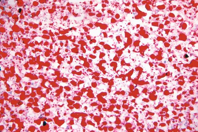
| 16:58, 19 August 2013 | IPLab2FattyChange8.jpg (file) |  |
78 KB | An oil red O stain for fat was performed on a frozen section of this liver tissue. The red droplets represent fat in the tissue which is typical of fatty degeneration in the liver. By using frozen sections the tissues do not have to be dehydrated throu... | 1 |
| 16:58, 19 August 2013 | IPLab2FattyChange7.jpg (file) |  |
65 KB | A high-power photomicrograph of the liver parenchyma shows that each individual liver cell is filled with a large, clear droplet which represents the space remaining after lipid was dissolved by the dehydration procedure used to embed the tissue. '''No... | 1 |
| 16:57, 19 August 2013 | IPLab2FattyChange6.jpg (file) |  |
67 KB | Another view at the same power illustrates the proliferation of bile ducts in the interlobular and perichordal regions (arrows). | 1 |
| 16:56, 19 August 2013 | IPLab2FattyChange5.jpg (file) |  |
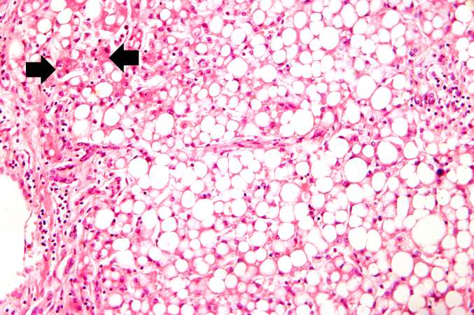
75 KB | This higher-power photomicrograph of the centrilobular area gives the appearance of fatty tissue, as indicated by many empty spaces. Very few normal liver cells can be seen in this slide. A few more normal-appearing hepatocytes are present at the left ... | 1 |
| 16:56, 19 August 2013 | IPLab2FattyChange4.jpg (file) |  |
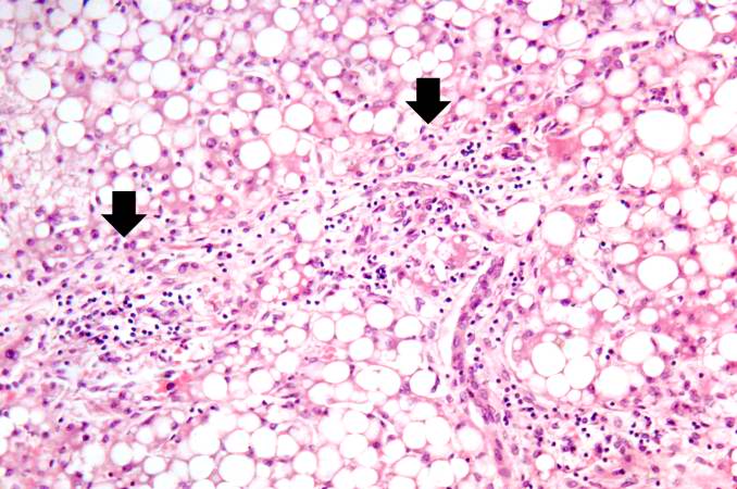
77 KB | A higher-power photomicrograph illustrates more clearly the inflammatory cells (arrows) around the portal areas. | 1 |
| 16:55, 19 August 2013 | IPLab2FattyChange3.jpg (file) |  |
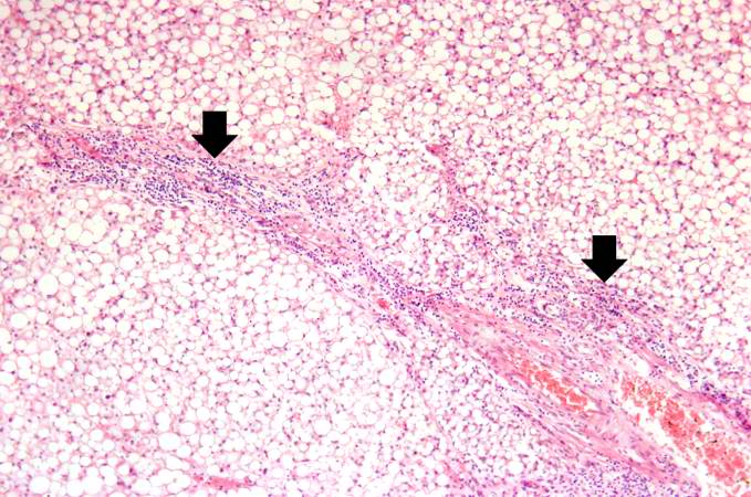
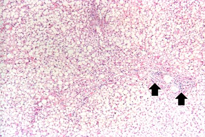
73 KB | Another low-power photomicrograph illustrates again the pale, washed-out appearance of this tissue. Notice the numerous holes throughout the tissue. There are accumulations of inflammatory cells (arrows) around portal tracts. | 1 |