IPLab:Lab 11:Ascariasis
Images
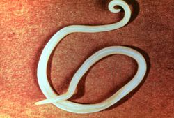
This is a photograph of an adult ascarid like the ones removed from the small bowel of this patient.
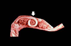
This is a photograph of an autopsy specimen from another case of ascariasis. The adult ascarid (arrow) can be seen in the section of small bowel from this patient.
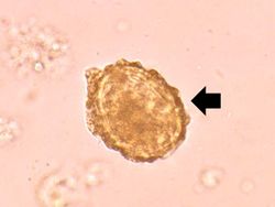
This is a high-power photomicrograph of a fecal specimen from this patient showing an ascarid egg (arrow).
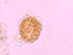
This is a higher-power photomicrograph of another ascarid egg.
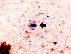
This high-power photomicrograph of the fecal specimen from this same patient shows an Entamoeba histolytica cyst (arrow).
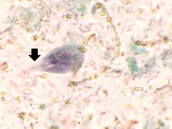
This high-power photomicrograph of the fecal specimen from this patient shows a Giardia lamblia trophozoite. Note the two nuclei and the tapered end (that goes out of the plane of focus) containing flagella (arrow).
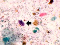
This high-power photomicrograph of the fecal specimen from this patient shows a Giardia lamblia cyst (arrow).


| |||||