IPLab:Lab 9:Bacterial Meningitis
Images
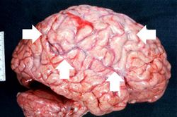
This gross photograph of the autopsy specimen from this case demonstrates the purulent exudate (arrows) in the leptomeninges.
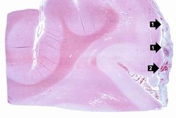
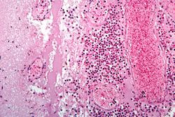
This is a low-power photomicrograph of brain section. Note the exudate (1) in the meninges and congestion of the vessels (2) in the leptomeninges.
This is a higher-power view of a congested blood vessel. Inflammatory exudate is present within the vessel and throughout the leptomeninges.
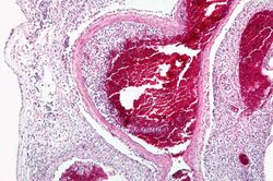
This higher-power photomicrograph of a sulcus shows the congested vessels and the inflammatory exudate in the leptomeninges.
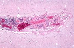
This is a higher-power photomicrograph of inflammatory exudate in a sulcus. The majority of cells in this exudate are neutrophils. There is also abundant fibrin (arrows) and red blood cells are present in the congested vessels.
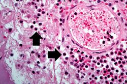
This is a high-power photomicrograph of a blood vessel from the previous image. The vessel is surrounded by neutrophils (arrows).
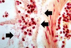
This is a high-power photomicrograph of exudate from the leptomeninges which has been Gram-stained. Note the Gram-negative bacteria (arrows) throughout this section.
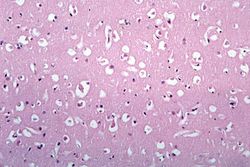
This photomicrograph of brain tissue demonstrates diffuse edema.


| |||||