IPLab:Lab 8:Rabies
Revision as of 02:41, 21 August 2013 by Seung Park (talk | contribs) (Created page with "== Images == <gallery heights="250px" widths="250px"> File:IPLab8Rabies1.jpg|This is a low-power photomicrograph of the hippocampus (arrow) from this case. File:IPLab8Rabies2...")
Images
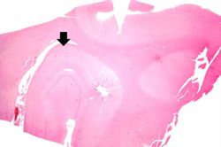
This is a low-power photomicrograph of the hippocampus (arrow) from this case.
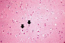
This is a medium-power photomicrograph of brain tissue exhibiting edema and evidence of shrunken, necrotic neurons (arrows).
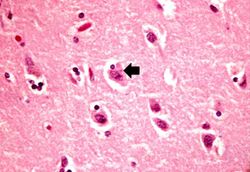
This is a higher-power photomicrograph of the shrunken neurons. One neuron appears to have an eosinophilic intracytoplasmic inclusion body (arrow).
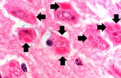
This is a high-power photomicrograph of neurons containing variably-sized intracytoplasmic eosinophilic inclusion bodies (arrows).
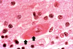
This is a high-power photomicrograph of a neuron surrounded by inflammatory cells (lymphocytes and microglia). This neuron has two intracytoplasmic eosinophilic inclusion bodies (arrows).


| |||||