File list
This special page shows all uploaded files.
| Date | Name | Thumbnail | Size | User | Description | Versions |
|---|---|---|---|---|---|---|
| 20:29, 9 January 2014 | CytologicallyYoursCoW20131118Cytology2.jpg (file) |  |
147 KB | Stephanie Simmons | 1 | |
| 20:28, 9 January 2014 | CytologicallyYoursCoW20131118Cytology1.jpg (file) |  |
64 KB | Stephanie Simmons | 1 | |
| 20:28, 9 January 2014 | CytologicallyYoursCoW20131118Biopsy3.jpg (file) |  |
165 KB | Stephanie Simmons | 1 | |
| 20:27, 9 January 2014 | CytologicallyYoursCoW20131118Biopsy2.jpg (file) |  |
162 KB | Stephanie Simmons | 1 | |
| 20:27, 9 January 2014 | CytologicallyYoursCoW20131111Radiology3.png (file) |  |
129 KB | Stephanie Simmons | 1 | |
| 20:27, 9 January 2014 | CytologicallyYoursCoW20131111Radiology2.png (file) |  |
114 KB | Stephanie Simmons | 1 | |
| 20:26, 9 January 2014 | CytologicallyYoursCoW20131111Radiology1.png (file) |  |
199 KB | Stephanie Simmons | 1 | |
| 20:26, 9 January 2014 | CytologicallyYoursCoW20131111Cytology6.jpg (file) |  |
80 KB | Stephanie Simmons | 1 | |
| 20:26, 9 January 2014 | CytologicallyYoursCoW20131111Cytology5.jpg (file) |  |
70 KB | Stephanie Simmons | 1 | |
| 20:25, 9 January 2014 | CytologicallyYoursCoW20131111Cytology4.jpg (file) |  |
77 KB | Stephanie Simmons | 1 | |
| 20:25, 9 January 2014 | CytologicallyYoursCoW20131111Cytology3.jpg (file) |  |
68 KB | Stephanie Simmons | 1 | |
| 20:22, 9 January 2014 | CytologicallyYoursCoW20131111Cytology2.jpg (file) |  |
87 KB | Stephanie Simmons | 1 | |
| 20:21, 9 January 2014 | CytologicallyYoursCoW20131111Cytology1.jpg (file) |  |
146 KB | Stephanie Simmons | 1 | |
| 20:21, 9 January 2014 | CytologicallyYoursCoW20131111Biopsy8.jpg (file) |  |
130 KB | Stephanie Simmons | 1 | |
| 20:21, 9 January 2014 | CytologicallyYoursCoW20131111Biopsy7.jpg (file) |  |
67 KB | Stephanie Simmons | 1 | |
| 20:21, 9 January 2014 | CytologicallyYoursCoW20131111Biopsy6.jpg (file) |  |
112 KB | Stephanie Simmons | 1 | |
| 20:20, 9 January 2014 | CytologicallyYoursCoW20131111Biopsy5.jpg (file) |  |
105 KB | Stephanie Simmons | 1 | |
| 20:20, 9 January 2014 | CytologicallyYoursCoW20131111Biopsy4.jpg (file) |  |
127 KB | Stephanie Simmons | 1 | |
| 20:19, 9 January 2014 | CytologicallyYoursCoW20131111Biopsy3.jpg (file) |  |
182 KB | Stephanie Simmons | 1 | |
| 20:19, 9 January 2014 | CytologicallyYoursCoW20131111Biopsy2.jpg (file) |  |
83 KB | Stephanie Simmons | 1 | |
| 20:19, 9 January 2014 | CytologicallyYoursCoW20131111Biopsy1.jpg (file) |  |
56 KB | Stephanie Simmons | 1 | |
| 22:27, 4 September 2013 | IPLab2Hypertrophy4.jpg (file) |  |
125 KB | Peter Anderson | 2 | |
| 22:26, 4 September 2013 | IPLab2Hypertrophy3.jpg (file) |  |
163 KB | Peter Anderson | 2 | |
| 22:24, 4 September 2013 | IPLab2Hypertrophy2.jpg (file) |  |
204 KB | Peter Anderson | 2 | |
| 06:06, 21 August 2013 | IPLab13Meningococcemia8.jpg (file) |  |
21 KB | Seung Park | This is a higher-power photomicrograph of a smear of cerebrospinal fluid taken at autopsy. Note the Gram-negative cocci in this smear, indicative of N. meningitidis. | 1 |
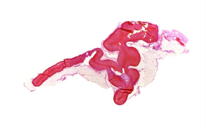
| 06:05, 21 August 2013 | IPLab13Meningococcemia7.jpg (file) |  |
34 KB | Seung Park | This higher-power photomicrograph of the adrenal gland from this case provides an example of hemorrhagic necrosis. | 1 |
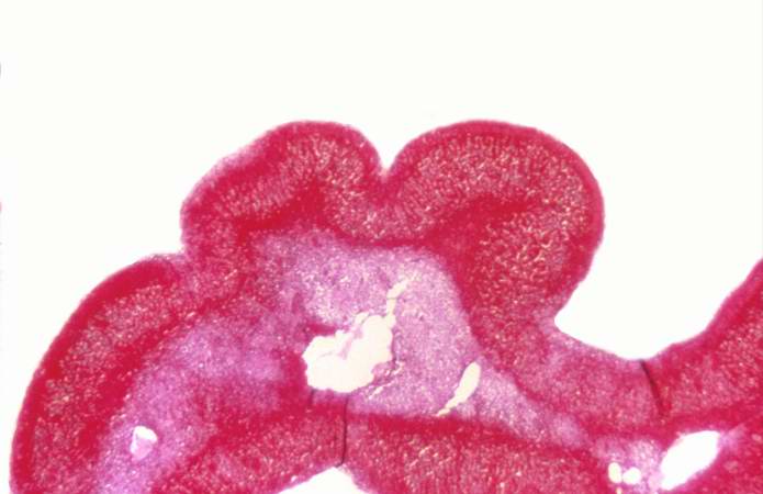
| 06:05, 21 August 2013 | IPLab13Meningococcemia6.jpg (file) |  |
20 KB | Seung Park | This is a low-power photomicrograph of the adrenal gland from this case. Note that the entire gland is hemorrhagic. | 2 |
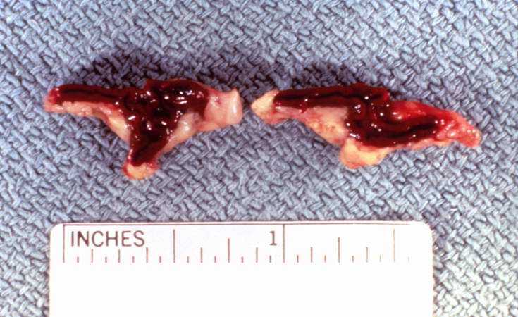
| 06:05, 21 August 2013 | IPLab13Meningococcemia5.jpg (file) |  |
56 KB | Seung Park | This is a gross photograph of cross sections through the adrenal glands from this case. Both adrenal glands are markedly hemorrhagic. | 1 |
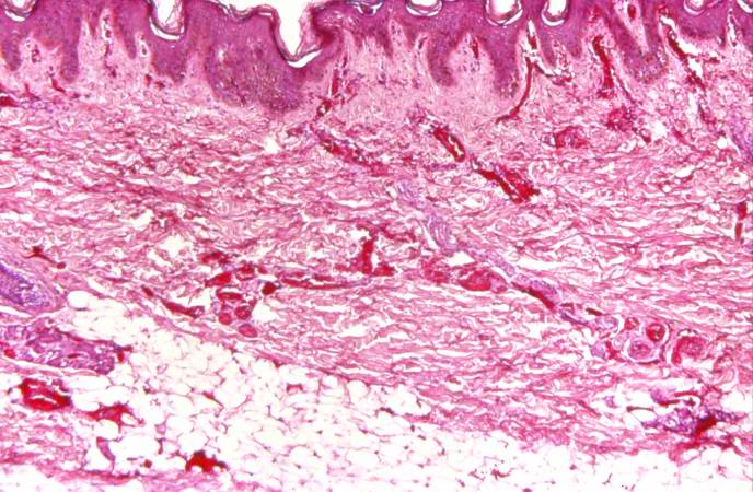
| 06:05, 21 August 2013 | IPLab13Meningococcemia4.jpg (file) |  |
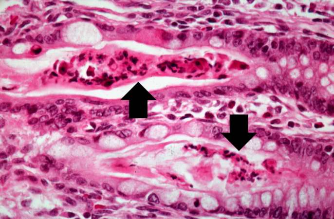
75 KB | Seung Park | This photomicrograph of the skin shows thrombi and fibrin clots in small vessels in the dermis. This is indicative of the endothelial damage caused by the Neisseria meningitidis endotoxin. This endotoxin-induced damage to the endothelium of small blood... | 1 |
| 06:05, 21 August 2013 | IPLab13Meningococcemia3.jpg (file) |  |
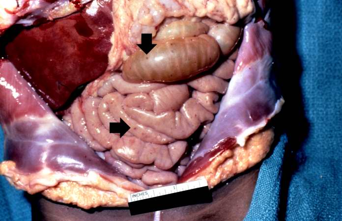
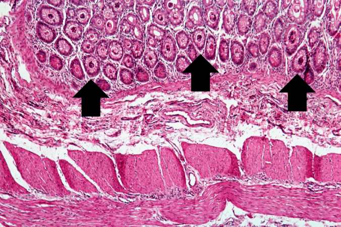
48 KB | Seung Park | In this gross photograph of the abdomen taken at the autopsy, there are petechial hemorrhages on the viscera (arrows). | 1 |
| 06:04, 21 August 2013 | IPLab13Meningococcemia2.jpg (file) |  |
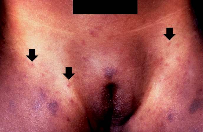
36 KB | Seung Park | This is a closer view of the inguinal region taken at autopsy. The areas of hemorrhage include purpura and petechiae (arrows). | 1 |
| 06:04, 21 August 2013 | IPLab13Meningococcemia1.jpg (file) |  |
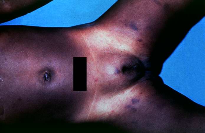
29 KB | Seung Park | In this gross photograph from the autopsy, note the areas of hemorrhage in the inguinal region. | 1 |
| 06:02, 21 August 2013 | IPLab13CF14.jpg (file) |  |
54 KB | Seung Park | Another high-power photomicrograph of intestine shows the vacuolated intestinal epithelial cells lining the crypts and necrotic debris and inspissated secretions within the crypts (arrows). | 1 |
| 06:02, 21 August 2013 | IPLab13CF13.jpg (file) |  |
71 KB | Seung Park | A higher-power photomicrograph of intestine shows the vacuolated intestinal epithelial cells lining the crypts and necrotic debris and inspissated secretions within the crypts (arrows). | 1 |
| 06:01, 21 August 2013 | IPLab13CF12.jpg (file) |  |
86 KB | Seung Park | This is a low-power photomicrograph from another section of the intestine. Saggital sections of the intestinal crypts show the crypts along their full length, extending to the mucosal surface. | 1 |
| 06:01, 21 August 2013 | IPLab13CF11.jpg (file) |  |
60 KB | Seung Park | This higher-power photomicrograph shows more clearly the eosinophilic debris (arrows) in the intestinal crypts. | 1 |
| 06:01, 21 August 2013 | IPLab13CF10.jpg (file) |  |
74 KB | Seung Park | This is a higher-power photomicrograph showing the eosinophilic debris in many of the intestinal crypts (arrows). | 1 |
| 06:01, 21 August 2013 | IPLab13CF9.jpg (file) |  |
87 KB | Seung Park | A higher-power photomicrograph shows the bottom of the intestinal crypts and the other normal layers of the intestine. Even at this magnification, accumulations of eosinophilic debris can be seen in many of the intestinal crypts (arrows). | 1 |
| 06:00, 21 August 2013 | IPLab13CF8.jpg (file) |  |
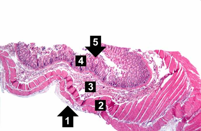
47 KB | Seung Park | This low-power photomicrograph of intestine shows the normal layers of the intestine, including the serosa (1), the muscularis (2), the submucosa (3), and the mucosal layer (4) with its deep mucosal crypts. There is yet another cystic space within the ... | 1 |
| 06:00, 21 August 2013 | IPLab13CF7.jpg (file) |  |
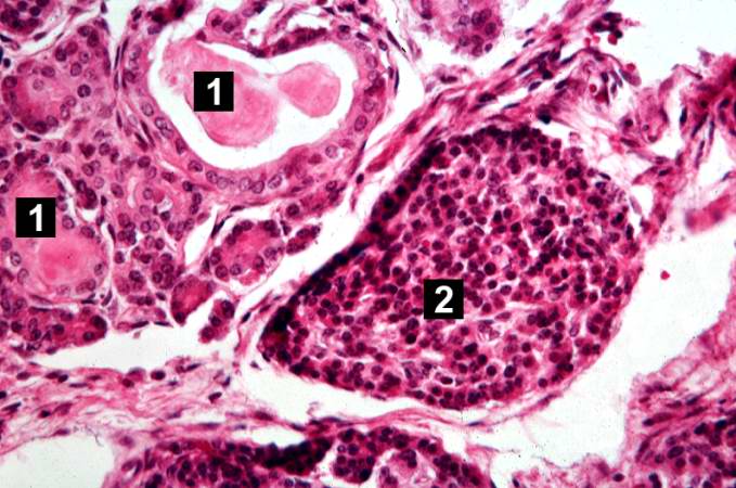
64 KB | Seung Park | This is another high-power photomicrograph showing cystic spaces (1) within the acinar pancreas and a normal islet of Langerhans (2). | 1 |
| 06:00, 21 August 2013 | IPLab13CF6.jpg (file) |  |
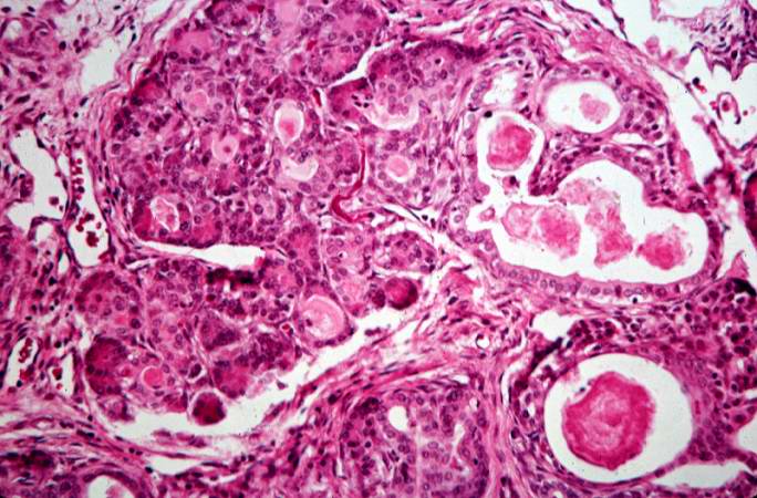
73 KB | Seung Park | This high-power photomicrograph shows more clearly these variably-sized cystic spaces within the acinar pancreas. | 1 |
| 06:00, 21 August 2013 | IPLab13CF5.jpg (file) |  |
74 KB | Seung Park | This higher-power photomicrograph shows a cystic space (1) within an acinar lobule. Islets of Langerhans (2) are also visible. | 1 |
| 06:00, 21 August 2013 | IPLab13CF4.jpg (file) |  |
85 KB | Seung Park | This higher-power photomicrograph of the pancreas shows interstitial tissue and the presence of small cystic spaces (1) within the acinar lobules. These spaces are filled with an eosinophilic proteinaceous material. The islets of Langerhans (2) are una... | 1 |
| 05:59, 21 August 2013 | IPLab13CF3.jpg (file) |  |
83 KB | Seung Park | This low-power photomicrograph of pancreas shows increased interstitial connective tissue resulting in accentuation of the lobular pattern. | 1 |
| 05:59, 21 August 2013 | IPLab13CF2.jpg (file) |  |
55 KB | Seung Park | This section of duodenum demonstrates dilation, loss of rugae, and areas of ulceration (arrows). | 1 |
| 05:59, 21 August 2013 | IPLab13CF1.jpg (file) |  |
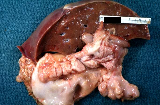
52 KB | Seung Park | This is a gross photograph of liver and pancreas from the autopsy. The pancreas is slightly smaller than normal and it has a mucous consistency. | 1 |
| 05:56, 21 August 2013 | IPLab13WT10.jpg (file) |  |
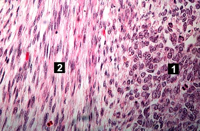
78 KB | Seung Park | This high-power photomicrograph shows the differences in cell morphology between the blastema (1) and the fibroblast type cells (2). | 1 |
| 05:56, 21 August 2013 | IPLab13WT9.jpg (file) |  |
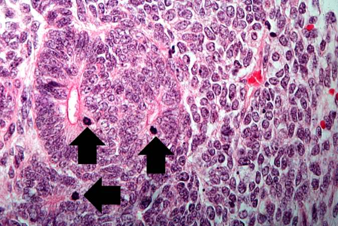
81 KB | Seung Park | This high-power photomicrograph demonstrates tubule formation within the blastema. Note the numerous mitotic figures (arrows). | 1 |
| 05:56, 21 August 2013 | IPLab13WT8.jpg (file) |  |
72 KB | Seung Park | This is a high-power photomicrograph of "tubule" formation within the blastema (arrows). | 1 |
| 05:55, 21 August 2013 | IPLab13WT7.jpg (file) |  |
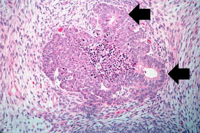
78 KB | Seung Park | This is another medium-power photomicrograph of the tumor. It demonstrates again the two cell types making up this neoplasm. The glands or "tubules" within the blastema are better developed in this section (arrows). | 1 |