File list
This special page shows all uploaded files.
| Date | Name | Thumbnail | Size | User | Description | Versions |
|---|---|---|---|---|---|---|
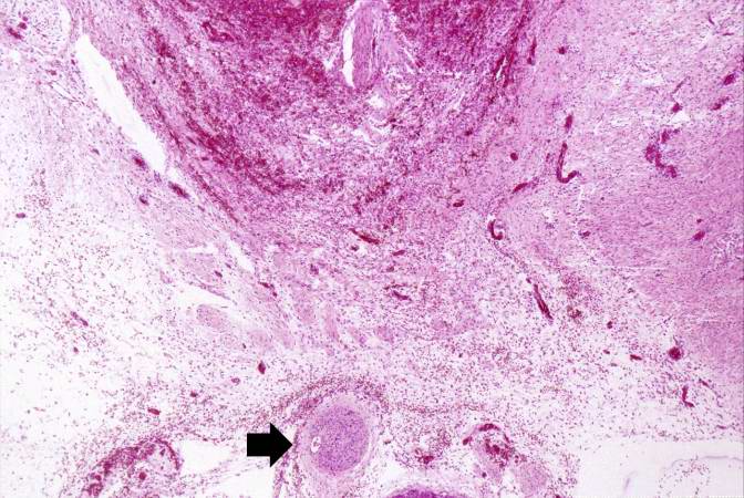
| 05:55, 21 August 2013 | IPLab13WT6.jpg (file) |  |
84 KB | Seung Park | This medium-power photomicrograph of tumor shows again the two cell types making up this neoplasm. There are regions within the blastema where the cells form glands or "tubules" (arrows). | 1 |
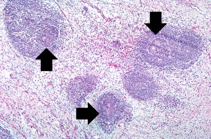
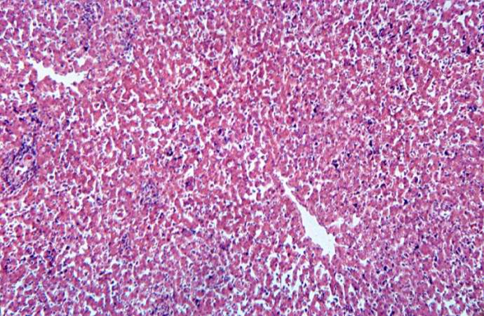
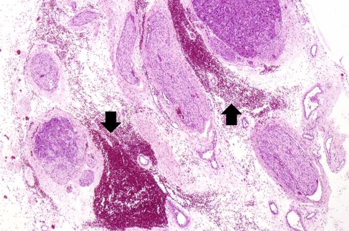
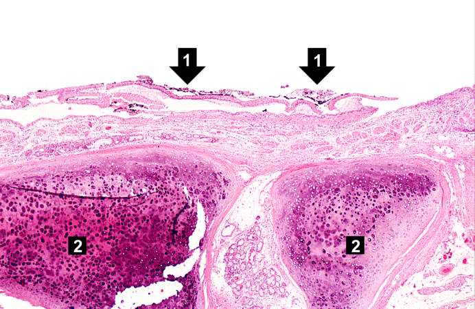
| 05:55, 21 August 2013 | IPLab13WT5.jpg (file) |  |
91 KB | Seung Park | This low-power photomicrograph of tumor shows the two cell types making up this neoplasm. The basophilic cellular component termed "blastema" (1) can be distinguished from less cellular eosinophilic areas with fibroblast-like cells (2). | 1 |
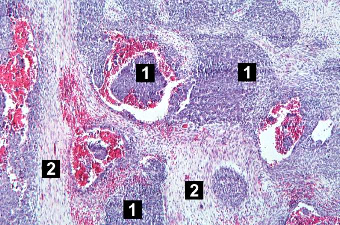
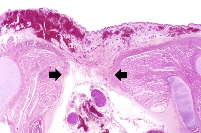
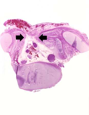
| 05:55, 21 August 2013 | IPLab13WT4.jpg (file) |  |
30 KB | Seung Park | This lowest-power view shows the tumor itself; no tissue is present that can be readily identified as normal kidney. There does appear to be a capsule surrounding the tumor. Eosinophilic bands are seen surrounding basophilic islands of cells. These cor... | 1 |
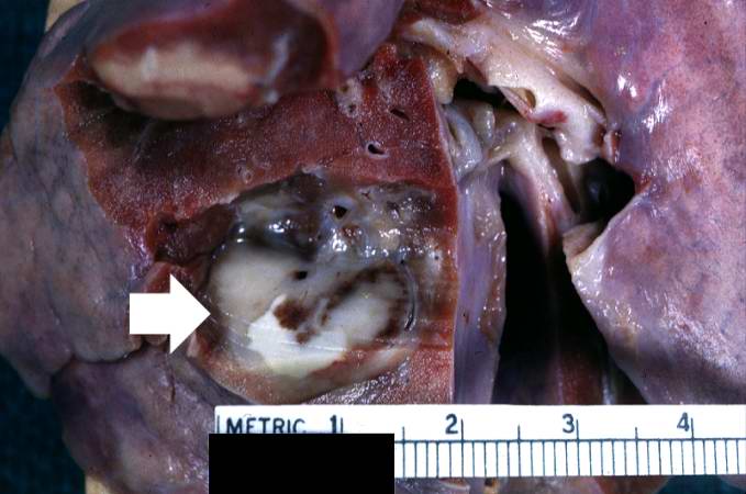
| 05:55, 21 August 2013 | IPLab13WT3.jpg (file) |  |
43 KB | Seung Park | This is a gross photograph of lung from this case demonstrating the metastatic tumor nodule (arrow). | 1 |
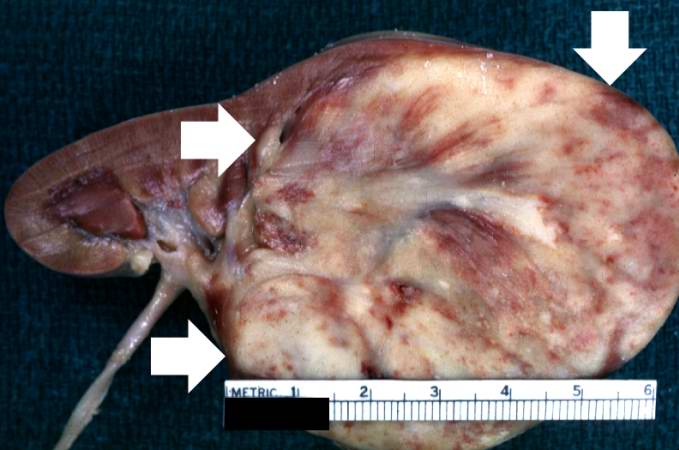
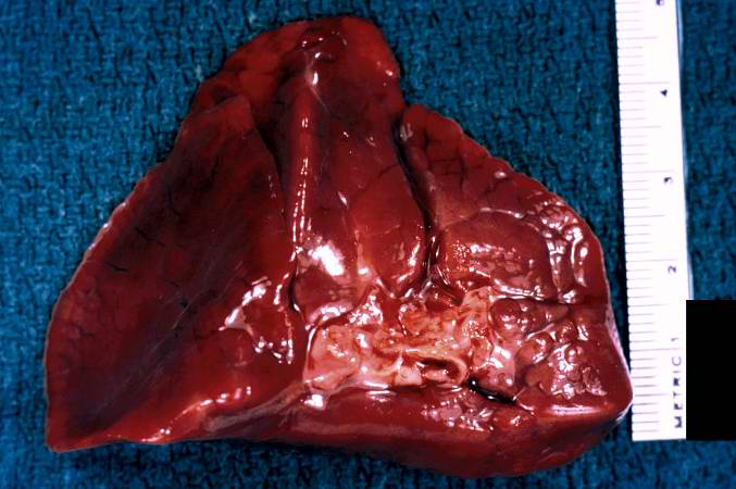
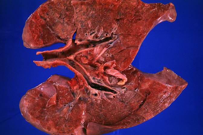
| 05:54, 21 August 2013 | IPLab13WT2.jpg (file) |  |
39 KB | Seung Park | This is a closer view of the kidney with Wilms' tumor (arrows). | 1 |
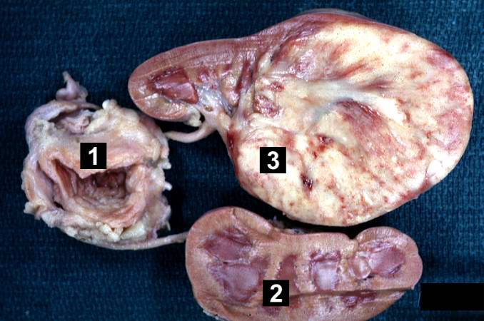
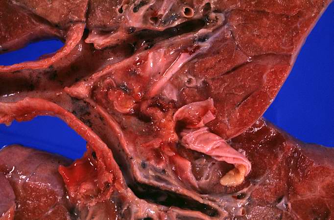
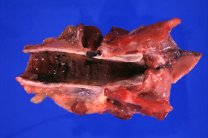
| 05:54, 21 August 2013 | IPLab13WT1.jpg (file) |  |
51 KB | Seung Park | This is a gross photograph of a bladder (1) to which are attached a normal kidney (2) and a kidney with Wilms' tumor (3). A large mass extends from the superior pole of the affected kidney. The renal capsule can be seen extending around this tumor. | 1 |
| 05:52, 21 August 2013 | IPLab13Hyaline9.jpg (file) |  |
78 KB | Seung Park | This higher-power photomicrograph shows more clearly the hyaline membranes (arrows) and the congestion in the interstitium. | 1 |
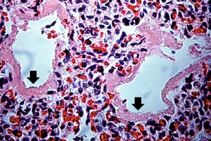
| 05:52, 21 August 2013 | IPLab13Hyaline8.jpg (file) |  |
90 KB | Seung Park | This medium-power photomicrograph shows the pink acellular homogeneous material lining the alveoli which comprises the hyaline membranes (arrows). The interstitium shows congestion, as in previous sections. | 1 |
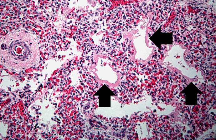
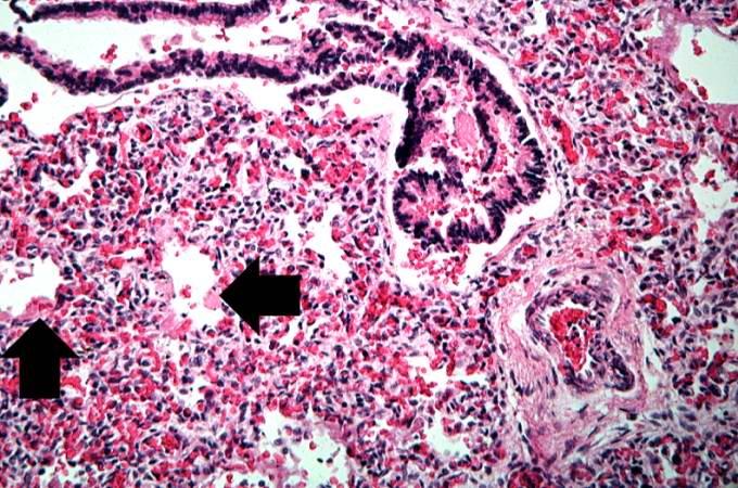
| 05:51, 21 August 2013 | IPLab13Hyaline7.jpg (file) |  |
93 KB | Seung Park | This high-power photomicrograph shows an airway with adjacent lung tissue. Some alveoli have hyaline membranes (arrows). There is severe congestion of the interstitium throughout this section. | 1 |
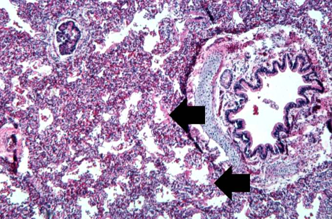
| 05:51, 21 August 2013 | IPLab13Hyaline6.jpg (file) |  |
85 KB | Seung Park | This is a medium-power photomicrograph showing a large bronchus with cartilage. Interstitial congestion with numerous red cells is apparent. Even at this magnification hyaline membranes (arrows) can be seen lining the alveoli. | 1 |
| 05:51, 21 August 2013 | IPLab13Hyaline5.jpg (file) |  |
78 KB | Seung Park | This low-power photomicrograph of lung demonstrates hypercellular pulmonary interstitium and small air spaces (as compared to adult lungs). | 1 |
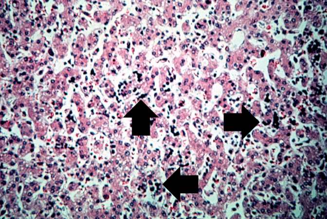
| 05:51, 21 August 2013 | IPLab13Hyaline4.jpg (file) |  |
81 KB | Seung Park | This high-power photomicrograph of liver shows more clearly the immature blood cell precursors (arrows) which represent extramedullary hematopoiesis of the liver. The liver is a normal site of fetal hematopoiesis and, for this stage of gestation, extra... | 1 |
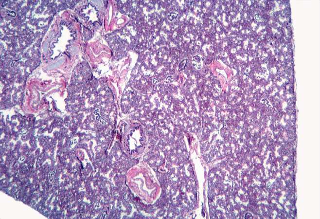
| 05:51, 21 August 2013 | IPLab13Hyaline3.jpg (file) |  |
89 KB | Seung Park | This is a low-power photomicrograph of liver which contains dark blue-stained cells in the hepatic sinusoids. These are immature blood cell precursors and this represents extramedullary hematopoiesis of the liver. | 1 |
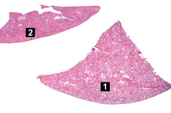
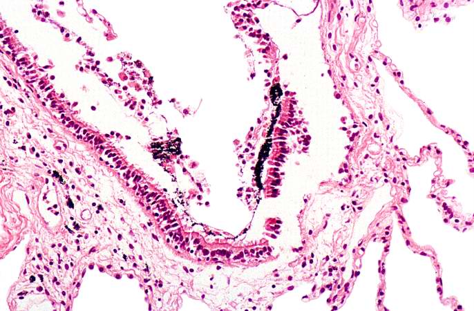
| 05:50, 21 August 2013 | IPLab13Hyaline2.jpg (file) |  |
36 KB | Seung Park | This is a low-power photomicrograph of a triangular-shaped section of lung (1) and an oblong section of liver (2). The lack of open air spaces in this neonatal lung indicates its immaturity. | 1 |
| 05:50, 21 August 2013 | IPLab13Hyaline1.jpg (file) |  |
46 KB | Seung Park | This is a gross photograph of lung demonstrating hyaline membrane disease and atelectasis. | 1 |
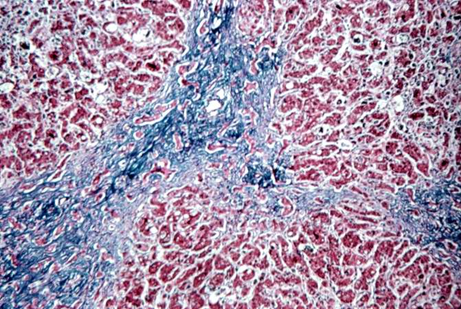
| 05:48, 21 August 2013 | IPLab13BiliaryAtresia5.jpg (file) |  |
87 KB | Seung Park | This is a medium-power photomicrograph of liver section stained with a trichrome stain to demonstrate the portal fibrosis. The fibrous connective tissue (collagen) stains blue. | 1 |
| 05:48, 21 August 2013 | IPLab13BiliaryAtresia4.jpg (file) |  |
72 KB | Seung Park | This is a high-power photomicrograph of fibrotic portal region with several bile ducts that contain inspissated bile (arrows). Adjacent hepatocytes also contain bile pigments. | 1 |
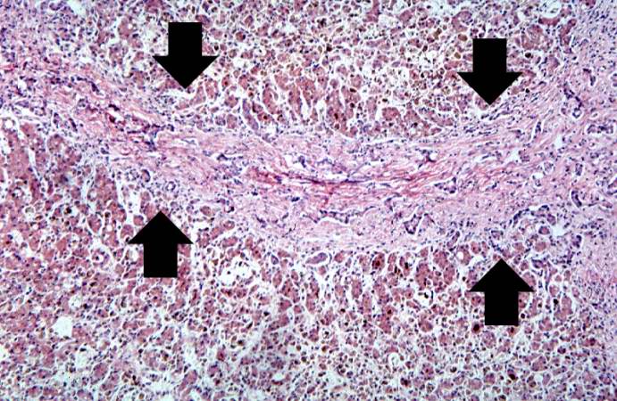
| 05:48, 21 August 2013 | IPLab13BiliaryAtresia3.jpg (file) |  |
82 KB | Seung Park | This high-power photomicrograph of fibrotic portal region demonstrates proliferation of the bile ducts (arrows). | 1 |
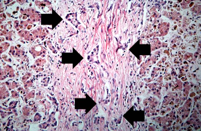
| 05:47, 21 August 2013 | IPLab13BiliaryAtresia2.jpg (file) |  |
95 KB | Seung Park | This medium-power photomicrograph of liver shows an area of portal fibrosis and bile duct proliferation (arrows). Adjacent to this fibrotic portal region, hepatocytes are seen separated by dilated sinusoids. Throughout this section are found accumulati... | 1 |
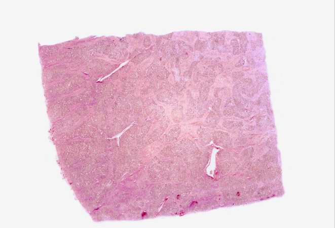
| 05:47, 21 August 2013 | IPLab13BiliaryAtresia1.jpg (file) |  |
30 KB | Seung Park | This is a low power photomicrograph of a section of liver. Even at this low magnification, areas of fibrosis can be appreciated. | 1 |
| 05:45, 21 August 2013 | IPLab13Myelomeningocele7.jpg (file) |  |
80 KB | Seung Park | This high-power photomicrograph of the spinal cord within the vertebral column shows the hemorrhage (arrows) in this region. | 1 |
| 05:45, 21 August 2013 | IPLab13Myelomeningocele6.jpg (file) |  |
77 KB | Seung Park | This is a high-power photomicrograph of the spinal cord (arrow) immediately beneath the area of hemorrhage. | 1 |
| 05:45, 21 August 2013 | IPLab13Myelomeningocele5.jpg (file) |  |
49 KB | Seung Park | This is a higher-power photomicrograph of one of the vertebral bodies from this case. The defect (arrows) in the vertebral body is seen more clearly. The spinal cord is disrupted and there are areas of hemorrhage in this region. | 1 |
| 05:45, 21 August 2013 | IPLab13Myelomeningocele4.jpg (file) |  |
16 KB | Seung Park | This is a low-power photomicrograph of one of the vertebral bodies from this case. In this section there are defects (arrows) in the vertebral body but the skin can be seen over the open vertebral canal. | 1 |
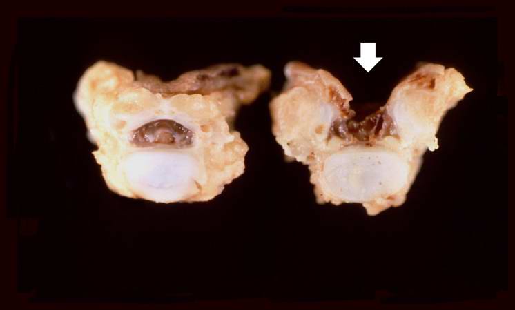
| 05:45, 21 August 2013 | IPLab13Myelomeningocele3.jpg (file) |  |
17 KB | Seung Park | This is a closer view of the previous gross photograph showing a normal lumbar vertebra from this case on the left. Once again, note the defect (arrow) in the vertebral body on the right due to failure of the vertebral column to close properly. | 1 |
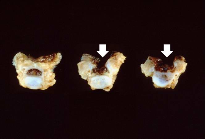
| 05:44, 21 August 2013 | IPLab13Myelomeningocele2.jpg (file) |  |
15 KB | Seung Park | This gross photograph shows consecutive lumbar vertebra from this case. Note the defect (arrows) in the two vertebral bodies on the right. This defect was caused by failure of the vertebral column to properly close. | 1 |
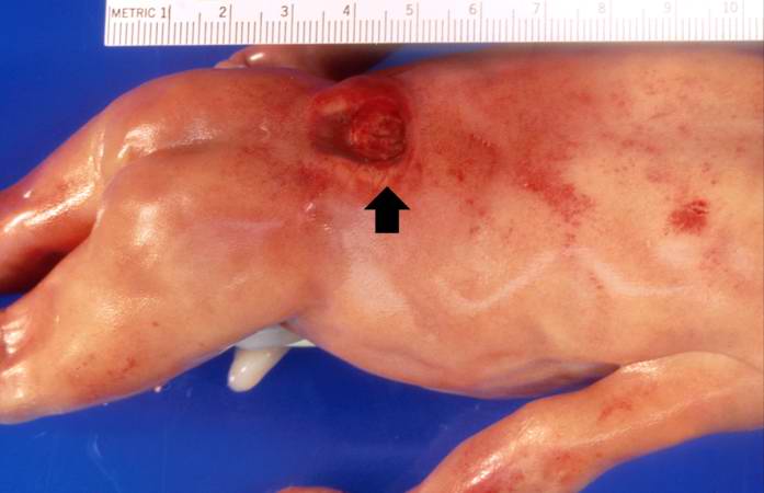
| 05:44, 21 August 2013 | IPLab13Myelomeningocele1.jpg (file) |  |
27 KB | Seung Park | This is a gross photograph of the fetus at autopsy. Note the defect in the lower lumbar region of the spinal column (arrow). The myelomeningocele can be seen protruding from this defect. | 1 |
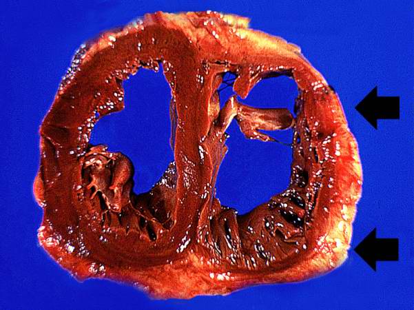
| 05:42, 21 August 2013 | IPLab12COPD5.jpg (file) |  |
50 KB | Seung Park | This gross photograph of the heart taken at autopsy demonstrates right ventricular hypertrophy and dilatation (arrows). | 1 |
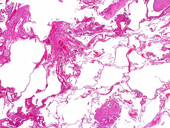
| 05:42, 21 August 2013 | IPLab12COPD4.jpg (file) |  |
68 KB | Seung Park | This higher-power photomicrograph of the lung shows more clearly the enlarged air spaces indicative of emphysematous change. | 1 |
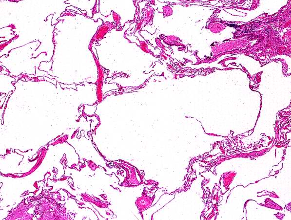
| 05:42, 21 August 2013 | IPLab12COPD3.jpg (file) |  |
73 KB | Seung Park | This low-power photomicrograph of the lung demonstrates the enlarged air spaces indicative of emphysematous change. | 1 |
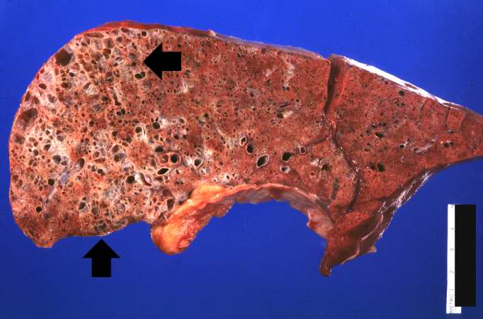
| 05:42, 21 August 2013 | IPLab12COPD2.jpg (file) |  |
70 KB | Seung Park | This is a higher-power view of the lung showing the emphysematous change. | 1 |
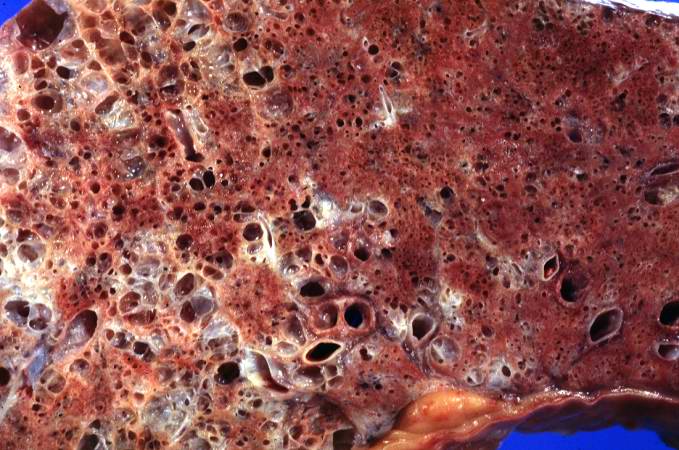
| 05:41, 21 August 2013 | IPLab12COPD1.jpg (file) |  |
45 KB | Seung Park | This gross photograph of lung taken at autopsy demonstrates the degree of emphysematous change (arrows). Also note that the rest of the lung is consolidated, indicating a pneumonia. | 1 |
| 05:40, 21 August 2013 | IPLab12Burns13.jpg (file) |  |
66 KB | Seung Park | 1 | |
| 05:39, 21 August 2013 | IPLab12Burns12.jpg (file) |  |
69 KB | Seung Park | This photomicrograph of the trachea shows sloughing of the tracheal epithelium and the black carbonaceous material contained therein (1). This degree of tracheal epithelial damage is indicative of severe inhalation injury. The tracheal cartilage rings ... | 1 |
| 05:39, 21 August 2013 | IPLab12Burns11.jpg (file) |  |
56 KB | Seung Park | This closer view of the lung shows the black carbonaceous material in the trachea as well as in the main stem bronchi. | 1 |
| 05:39, 21 August 2013 | IPLab12Burns10.jpg (file) |  |
45 KB | Seung Park | This photograph of the lung again shows the black carbonaceous material in the trachea as well as in the main stem bronchi. The lungs are mildly congested and hyperemic. The patient lived for less than 8 hours after the burn injury so there was not eno... | 1 |
| 05:39, 21 August 2013 | IPLab12Burns9.jpg (file) |  |
34 KB | Seung Park | This photograph demonstrates the black carbonaceous material in the trachea. | 1 |
| 05:38, 21 August 2013 | IPLab12Burns8.jpg (file) |  |
61 KB | Seung Park | This medium-power photomicrograph of the burned skin shows the mild damage to the superficial layers of the epidermis. | 1 |
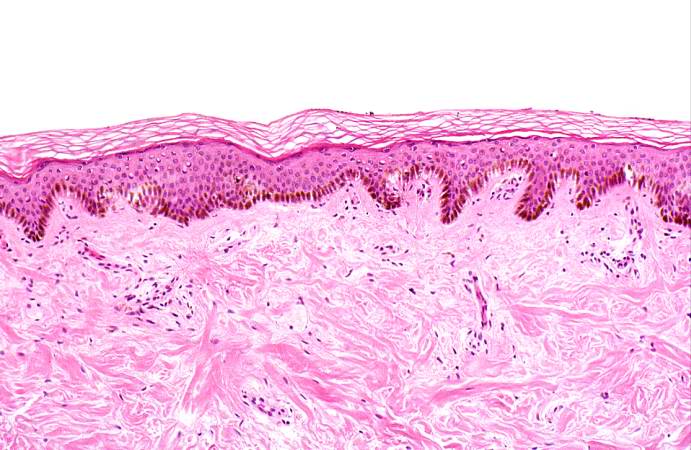
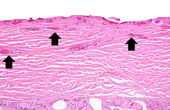
| 05:38, 21 August 2013 | IPLab12Burns7.jpg (file) |  |
68 KB | Seung Park | This photomicrograph of the burned skin depicts an area of first degree burn. Note that there are no blisters and no damage to the dermis. There is mild damage to the superficial epidermis and some hyperemia (arrow). | 1 |
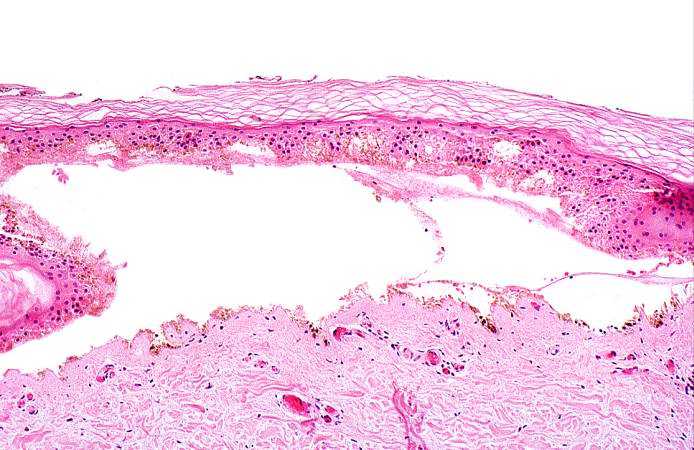
| 05:38, 21 August 2013 | IPLab12Burns6.jpg (file) |  |
63 KB | Seung Park | This high-power photomicrograph of the burned skin shows the blister and the thrombosed vessels in the dermis. | 1 |
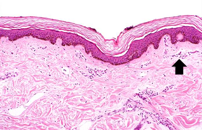
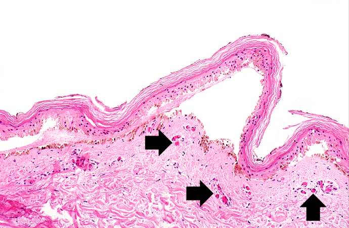
| 05:38, 21 August 2013 | IPLab12Burns5.jpg (file) |  |
56 KB | Seung Park | This medium-power photomicrograph of the burned skin demonstrates blister formation. The vessels in the dermis are congested (arrows). | 1 |
| 05:37, 21 August 2013 | IPLab12Burns4.jpg (file) |  |
68 KB | Seung Park | This high-power photomicrograph shows the denuded surface of the skin with thrombosed blood vessels (arrows) and necrosis of the dermis. | 1 |
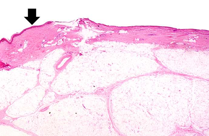
| 05:37, 21 August 2013 | IPLab12Burns3.jpg (file) |  |
44 KB | Seung Park | This photomicrograph of the burned skin depicts an area of third degree burn. There is some residual epidermis (arrow) but in the majority of this section the epidermis is gone. Also note the severe subcutaneous edema. | 1 |
| 05:37, 21 August 2013 | IPLab12Burns2.jpg (file) |  |
34 KB | Seung Park | This closer view shows the burned skin peeling off to expose the underlying tissue. The various depths of the burn can be appreciated by the color and character of the underlying tissue. | 1 |
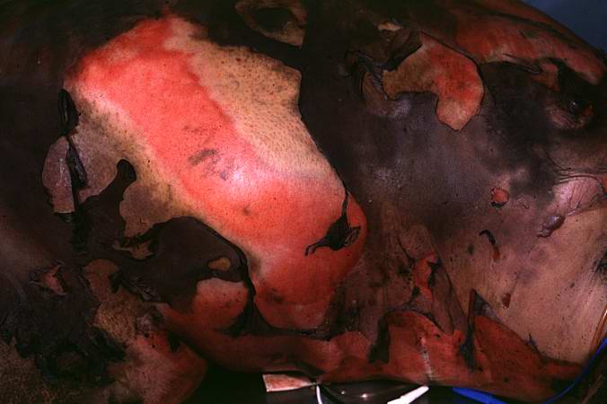
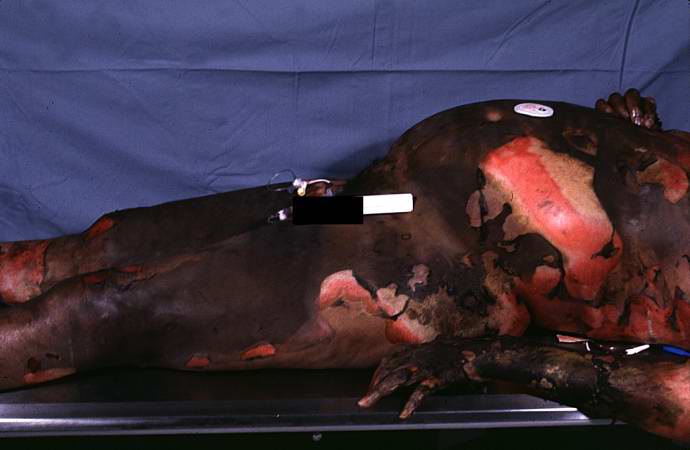
| 05:37, 21 August 2013 | IPLab12Burns1.jpg (file) |  |
30 KB | Seung Park | This photograph taken at autopsy demonstrates the severity of the surface burns on this patient. | 1 |
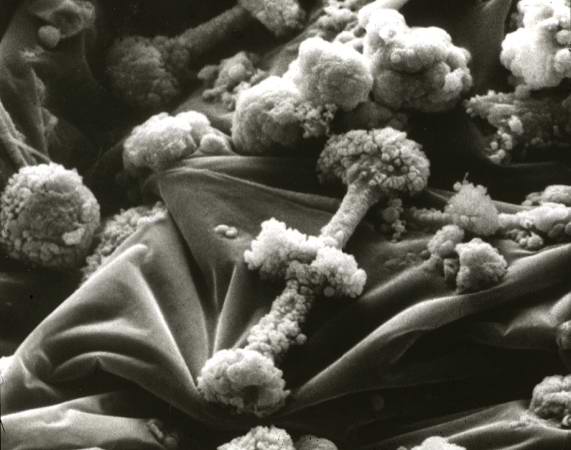
| 05:34, 21 August 2013 | IPLab12Mesothelioma11.jpg (file) |  |
35 KB | Seung Park | Scanning electron micrograph of asbestos bodies. Note the rough surface and the beaded appearance caused by the material adhering to the surface of the asbestos fiber. | 1 |
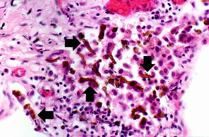
| 05:34, 21 August 2013 | IPLab12Mesothelioma10.jpg (file) |  |
81 KB | Seung Park | Another high-power photomicrograph of the brown asbestos bodies showing the characteristic beaded appearance (arrows). Tumor cells are evident adjacent to the asbestos bodies. | 1 |
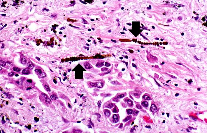
| 05:34, 21 August 2013 | IPLab12Mesothelioma9.jpg (file) |  |
64 KB | Seung Park | This is a high-power photomicrograph of the brown asbestos bodies showing the characteristic beaded appearance (arrows). | 1 |
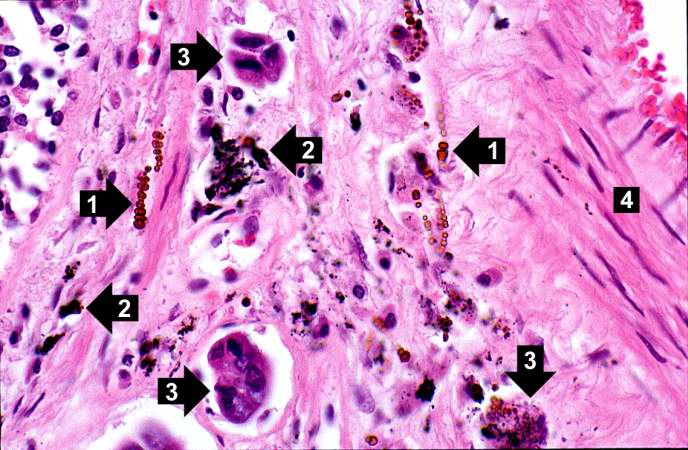
| 05:33, 21 August 2013 | IPLab12Mesothelioma8.jpg (file) |  |
68 KB | Seung Park | This high-power photomicrograph shows brown asbestos bodies (1), accumulations of anthracotic pigment (2), and tumor cells (3) all adjacent to a blood vessel (4). | 1 |
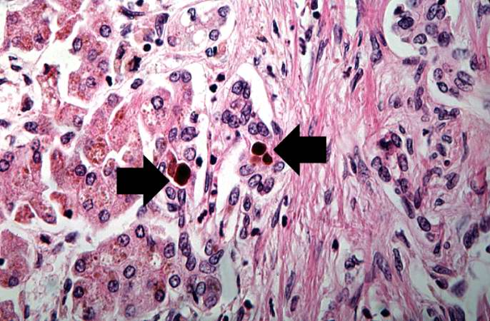
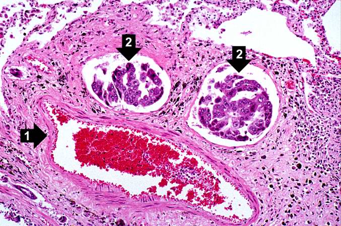
| 05:33, 21 August 2013 | IPLab12Mesothelioma7.jpg (file) |  |
100 KB | Seung Park | In this higher-power photomicrograph there is a blood vessel (1) and adjacent lymphatics that contain tumor cells (2). There are also accumulations of brown material adjacent to these vessels. | 1 |